
Page 195 - Fundamentals of Light Microscopy and Electronic Imaging
P. 195
178 FLUORESCENCE MICROSCOPY
10 µm
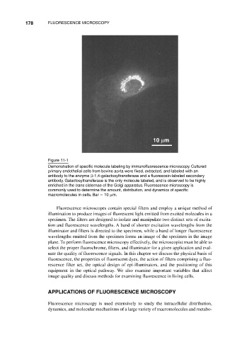
Figure 11-1
Demonstration of specific molecule labeling by immunofluorescence microscopy. Cultured
primary endothelial cells from bovine aorta were fixed, extracted, and labeled with an
antibody to the enzyme -1,4-galactosyltransferase and a fluorescein-labeled secondary
antibody. Galactosyltransferase is the only molecule labeled, and is observed to be highly
enriched in the trans cisternae of the Golgi apparatus. Fluorescence microscopy is
commonly used to determine the amount, distribution, and dynamics of specific
macromolecules in cells. Bar 10 m.
Fluorescence microscopes contain special filters and employ a unique method of
illumination to produce images of fluorescent light emitted from excited molecules in a
specimen. The filters are designed to isolate and manipulate two distinct sets of excita-
tion and fluorescence wavelengths. A band of shorter excitation wavelengths from the
illuminator and filters is directed to the specimen, while a band of longer fluorescence
wavelengths emitted from the specimen forms an image of the specimen in the image
plane. To perform fluorescence microscopy effectively, the microscopist must be able to
select the proper fluorochrome, filters, and illuminator for a given application and eval-
uate the quality of fluorescence signals. In this chapter we discuss the physical basis of
fluorescence, the properties of fluorescent dyes, the action of filters comprising a fluo-
rescence filter set, the optical design of epi-illuminators, and the positioning of this
equipment in the optical pathway. We also examine important variables that affect
image quality and discuss methods for examining fluorescence in living cells.
APPLICATIONS OF FLUORESCENCE MICROSCOPY
Fluorescence microscopy is used extensively to study the intracellular distribution,
dynamics, and molecular mechanisms of a large variety of macromolecules and metabo-