
Page 23 - Fundamentals of Light Microscopy and Electronic Imaging
P. 23
6 FUNDAMENTALS OF LIGHT MICROSCOPY
Conjugate Conjugate
field planes aperture planes
4 Retina Eye 4 Iris diaphragm
of eye
Eyepiece
Field stop of
3 Intermediate
eyepiece
image
3 Back focal
Objective lens plane of
2 Object plane Stage objective
Condenser lens
2 Front focal
plane of
condenser
1 Field stop
diaphragm Collector lens
Lamp 1 Lamp filament
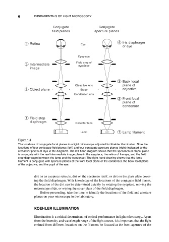
Figure 1-4
The locations of conjugate focal planes in a light microscope adjusted for Koehler illumination. Note the
locations of four conjugate field planes (left) and four conjugate aperture planes (right) indicated by the
crossover points of rays in the diagrams. The left-hand diagram shows that the specimen or object plane
is conjugate with the real intermediate image plane in the eyepiece, the retina of the eye, and the field
stop diaphragm between the lamp and the condenser. The right-hand drawing shows that the lamp
filament is conjugate with aperture planes at the front focal plane of the condenser, the back focal plane
of the objective, and the pupil of the eye.
dirt on an eyepiece reticule, dirt on the specimen itself, or dirt on the glass plate cover-
ing the field diaphragm. With knowledge of the locations of the conjugate field planes,
the location of the dirt can be determined quickly by rotating the eyepiece, moving the
microscope slide, or wiping the cover plate of the field diaphragm.
Before proceeding, take the time to identify the locations of the field and aperture
planes on your microscope in the laboratory.
KOEHLER ILLUMINATION
Illumination is a critical determinant of optical performance in light microscopy. Apart
from the intensity and wavelength range of the light source, it is important that the light
emitted from different locations on the filament be focused at the front aperture of the