
Page 19 - Fundamentals of Light Microscopy and Electronic Imaging
P. 19
2 FUNDAMENTALS OF LIGHT MICROSCOPY
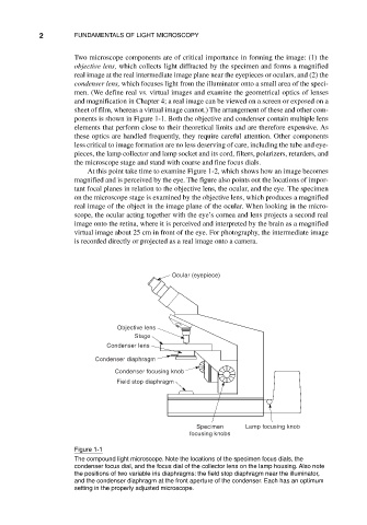
Two microscope components are of critical importance in forming the image: (1) the
objective lens, which collects light diffracted by the specimen and forms a magnified
real image at the real intermediate image plane near the eyepieces or oculars, and (2) the
condenser lens, which focuses light from the illuminator onto a small area of the speci-
men. (We define real vs. virtual images and examine the geometrical optics of lenses
and magnification in Chapter 4; a real image can be viewed on a screen or exposed on a
sheet of film, whereas a virtual image cannot.) The arrangement of these and other com-
ponents is shown in Figure 1-1. Both the objective and condenser contain multiple lens
elements that perform close to their theoretical limits and are therefore expensive. As
these optics are handled frequently, they require careful attention. Other components
less critical to image formation are no less deserving of care, including the tube and eye-
pieces, the lamp collector and lamp socket and its cord, filters, polarizers, retarders, and
the microscope stage and stand with coarse and fine focus dials.
At this point take time to examine Figure 1-2, which shows how an image becomes
magnified and is perceived by the eye. The figure also points out the locations of impor-
tant focal planes in relation to the objective lens, the ocular, and the eye. The specimen
on the microscope stage is examined by the objective lens, which produces a magnified
real image of the object in the image plane of the ocular. When looking in the micro-
scope, the ocular acting together with the eye’s cornea and lens projects a second real
image onto the retina, where it is perceived and interpreted by the brain as a magnified
virtual image about 25 cm in front of the eye. For photography, the intermediate image
is recorded directly or projected as a real image onto a camera.
Ocular (eyepiece)
Objective lens
Stage
Condenser lens
Condenser diaphragm
Condenser focusing knob
Field stop diaphragm
Specimen Lamp focusing knob
focusing knobs
Figure 1-1
The compound light microscope. Note the locations of the specimen focus dials, the
condenser focus dial, and the focus dial of the collector lens on the lamp housing. Also note
the positions of two variable iris diaphragms: the field stop diaphragm near the illuminator,
and the condenser diaphragm at the front aperture of the condenser. Each has an optimum
setting in the properly adjusted microscope.