
Page 238 - Fundamentals of Light Microscopy and Electronic Imaging
P. 238
ELECTRONIC ADJUSTMENTS AND CONSIDERATIONS 221
pling factor may need to be as high as 3–4 (instead of 2) to maintain the resolution pro-
vided by the objective.
Laser Selection and Laser Intensity
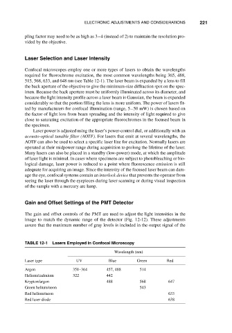
Confocal microscopes employ one or more types of lasers to obtain the wavelengths
required for fluorochrome excitation, the most common wavelengths being 365, 488,
515, 568, 633, and 648 nm (see Table 12-1). The laser beam is expanded by a lens to fill
the back aperture of the objective to give the minimum-size diffraction spot on the spec-
imen. Because the back aperture must be uniformly illuminated across its diameter, and
because the light intensity profile across a laser beam is Gaussian, the beam is expanded
considerably so that the portion filling the lens is more uniform. The power of lasers fit-
ted by manufacturers for confocal illumination (range, 5–50 mW) is chosen based on
the factor of light loss from beam spreading and the intensity of light required to give
close to saturating excitation of the appropriate fluorochromes in the focused beam in
the specimen.
Laser power is adjusted using the laser’s power-control dial, or additionally with an
acousto-optical tunable filter (AOTF). For lasers that emit at several wavelengths, the
AOTF can also be used to select a specific laser line for excitation. Normally lasers are
operated at their midpower range during acquisition to prolong the lifetime of the laser.
Many lasers can also be placed in a standby (low-power) mode, at which the amplitude
of laser light is minimal. In cases where specimens are subject to photobleaching or bio-
logical damage, laser power is reduced to a point where fluorescence emission is still
adequate for acquiring an image. Since the intensity of the focused laser beam can dam-
age the eye, confocal systems contain an interlock device that prevents the operator from
seeing the laser through the eyepieces during laser scanning or during visual inspection
of the sample with a mercury arc lamp.
Gain and Offset Settings of the PMT Detector
The gain and offset controls of the PMT are used to adjust the light intensities in the
image to match the dynamic range of the detector (Fig. 12-12). These adjustments
assure that the maximum number of gray levels is included in the output signal of the
TABLE 12-1 Lasers Employed in Confocal Microscopy
Wavelength (nm)
Laser type UV Blue Green Red
Argon 351–364 457, 488 514
Helium/cadmium 322 442
Krypton/argon 488 568 647
Green helium/neon 543
Red helium/neon 633
Red laser diode 638