
Page 30 - Fundamentals of Light Microscopy and Electronic Imaging
P. 30
PRECAUTIONS FOR HANDLING OPTICAL EQUIPMENT 13
Eyepiece Reticule disk Stage
for eyepiece micrometer
0.0 0.1 0.2 0.3 0.4
0 1 2 3 4 5 6 7
Overlapping reticule and micrometer scales
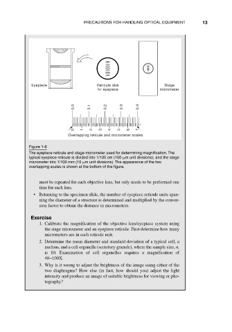
Figure 1-6
The eyepiece reticule and stage micrometer used for determining magnification. The
typical eyepiece reticule is divided into 1/100 cm (100 m unit divisions), and the stage
micrometer into 1/100 mm (10 m unit divisions). The appearance of the two
overlapping scales is shown at the bottom of the figure.
must be repeated for each objective lens, but only needs to be performed one
time for each lens.
• Returning to the specimen slide, the number of eyepiece reticule units span-
ning the diameter of a structure is determined and multiplied by the conver-
sion factor to obtain the distance in micrometers.
Exercise
1. Calibrate the magnification of the objective lens/eyepiece system using
the stage micrometer and an eyepiece reticule. First determine how many
micrometers are in each reticule unit.
2. Determine the mean diameter and standard deviation of a typical cell, a
nucleus, and a cell organelle (secretory granule), where the sample size, n,
is 10. Examination of cell organelles requires a magnification of
40–100X.
3. Why is it wrong to adjust the brightness of the image using either of the
two diaphragms? How else (in fact, how should you) adjust the light
intensity and produce an image of suitable brightness for viewing or pho-
tography?