
Page 35 - Fundamentals of Light Microscopy and Electronic Imaging
P. 35
18 LIGHT AND COLOR
Resolution
limit Wavelength class Size references
1mm
Radio
Human eye 100 m
Epithelial cells
Infrared 10 m
Red cells
Bacteria
1 m
Light Visible
microscope Mycoplasma
100 nm
Ultra-
violet Viruses
10 nm
Proteins
- and
x-rays 1 nm Amino acids
Electron
microscope 0.1 nm Atoms
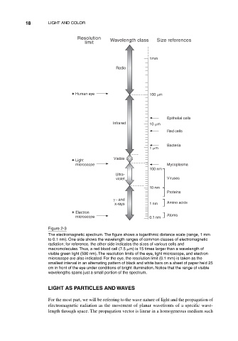
Figure 2-3
The electromagnetic spectrum. The figure shows a logarithmic distance scale (range, 1 mm
to 0.1 nm). One side shows the wavelength ranges of common classes of electromagnetic
radiation; for reference, the other side indicates the sizes of various cells and
macromolecules. Thus, a red blood cell (7.5 m) is 15 times larger than a wavelength of
visible green light (500 nm). The resolution limits of the eye, light microscope, and electron
microscope are also indicated. For the eye, the resolution limit (0.1 mm) is taken as the
smallest interval in an alternating pattern of black and white bars on a sheet of paper held 25
cm in front of the eye under conditions of bright illumination. Notice that the range of visible
wavelengths spans just a small portion of the spectrum.
LIGHT AS PARTICLES AND WAVES
For the most part, we will be referring to the wave nature of light and the propagation of
electromagnetic radiation as the movement of planar wavefronts of a specific wave-
length through space. The propagation vector is linear in a homogeneous medium such