
Page 36 - Fundamentals of Light Microscopy and Electronic Imaging
P. 36
LIGHT AS PARTICLES AND WAVES 19
as air or glass or in a vacuum. The relatively narrow spectrum of photon energies (and
corresponding frequencies) we experience as light is capable of exciting the visual pig-
ments in the rod and cone cells in the retina and corresponds to wavelengths ranging
from 400 nm (violet) to 750 nm (red). As shown in Figure 2-4, we depict light in vari-
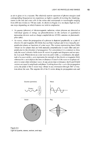
ous ways depending on which features we wish to emphasize:
• As quanta (photons) of electromagnetic radiation, where photons are detected as
individual quanta of energy (as photoelectrons) on the surfaces of quantitative
measuring devices such as charge-coupled device (CCD) cameras or photomulti-
plier tubes.
• As waves, where the propagation of a photon is depicted graphically as a pair of
electric (E) and magnetic (B) fields that oscillate in phase and in two mutually per-
pendicular planes as functions of a sine wave. The vectors representing these fields
vibrate in two planes that are both mutually perpendicular to each other and per-
pendicular to the direction of propagation. For convenience it is common to show
only the wave’s electric field vector (E vector) in graphs and diagrams and not spec-
ify it as such. When shown as a sine wave on a plot with x, y coordinates, the ampli-
tude of a wave on the y-axis represents the strength of the electric or magnetic field,
whereas the x-axis depicts the time or distance of travel of the wave or its phase rel-
ative to some other reference wave. At any given time or distance, the E and B field
vectors are equal in amplitude and phase. Looking down the x-axis (the propagation
axis), the plane of the E vector may vibrate in any orientation through 360° of rota-
tion about the axis. The angular tilt of the E vector along its propagation axis and
Quanta (particles) Wave
Photon
Vector Ray
Figure 2-4
Light as quanta, waves, vectors, and rays.