
Page 41 - Fundamentals of Light Microscopy and Electronic Imaging
P. 41
24 LIGHT AND COLOR
160 Rods
Number (thousands per mm 2 ) 80 Rods Blind spot
120
40
60°
80° 60° Cones 20° 0 20° 40° Cones 80° 100°
40°
(temporal side) (nasal side)
Angle from fovea
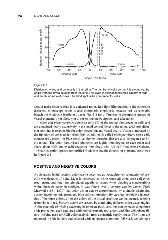
Figure 2-7
2
Distribution of rod and cone cells in the retina. The number of cells per mm is plotted vs. the
angle from the fovea as seen from the lens. The fovea is distinct in having a paucity of rods
and an abundance of cones. The blind spot lacks photoreceptor cells.
should make observations in a darkened room. Red light illumination in the otherwise
darkened microscope room is also commonly employed, because red wavelengths
bleach the rhodopsin inefficiently (see Fig. 2-8 for differences in absorption spectra of
visual pigments), yet allow you to see to operate equipment and take notes.
Cone cell photoreceptors comprise only 5% of the retinal photoreceptor cells and
are contained nearly exclusively in the small central fovea of the retina, a 0.5 mm diam-
eter spot that is responsible for color perception and visual acuity. Vision dominated by
the function of cones under bright light conditions is called photopic vision. Cone cells
contain red-, green-, or blue-sensitive pigment proteins that are also conjugated to 11-
cis-retinal. The color photovisual pigments are highly homologous to each other and
share about 40% amino acid sequence homology with rod cell rhodopsin (Nathans,
1984). Absorption spectra for purified rhodopsin and the three color pigments are shown
in Figure 2-8.
POSITIVE AND NEGATIVE COLORS
As discussed in this section, color can be described as the addition or subtraction of spe-
cific wavelengths of light. Light is perceived as white when all three cone cell types
(red, green, and blue) are stimulated equally as occurs when viewing a nonabsorbing
white sheet of paper in sunlight. It was found over a century ago by James Clerk
Maxwell (1831–1879) that color vision can be approximated by a simple tristimulus
system involving red, green, and blue color stimulation. By varying the relative intensi-
ties of the three colors, all of the colors of the visual spectrum can be created, ranging
from violet to red. Positive colors are created by combining different color wavelengths.
A fine example of mixing wavelengths to create positive colors can be made using three
slide projectors, each equipped with monochromatic red, green, and blue cellophane fil-
ters (the kind used for RGB color analysis) from a scientific supply house. The filters are
mounted in slide holders and covered with an opaque aluminum foil mask containing a