
Page 44 - Fundamentals of Light Microscopy and Electronic Imaging
P. 44
POSITIVE AND NEGATIVE COLORS 27
1st order
Spectrum
Lens spot
(white)
R
O
Y
G
B
I
V 0th order
spot
(white)
Screen
Continuous Infrared Slit
light filter
source
Diffraction grating
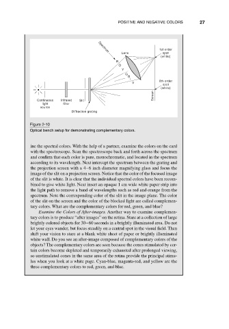
Figure 2-10
Optical bench setup for demonstrating complementary colors.
ine the spectral colors. With the help of a partner, examine the colors on the card
with the spectroscope. Scan the spectroscope back and forth across the spectrum
and confirm that each color is pure, monochromatic, and located in the spectrum
according to its wavelength. Next intercept the spectrum between the grating and
the projection screen with a 4–6 inch diameter magnifying glass and focus the
image of the slit on a projection screen. Notice that the color of the focused image
of the slit is white. It is clear that the individual spectral colors have been recom-
bined to give white light. Next insert an opaque 1 cm wide white paper strip into
the light path to remove a band of wavelengths such as red and orange from the
spectrum. Note the corresponding color of the slit in the image plane. The color
of the slit on the screen and the color of the blocked light are called complemen-
tary colors. What are the complementary colors for red, green, and blue?
Examine the Colors of After-images. Another way to examine complemen-
tary colors is to produce “after images” on the retina. Stare at a collection of large
brightly colored objects for 30–60 seconds in a brightly illuminated area. Do not
let your eyes wander, but focus steadily on a central spot in the visual field. Then
shift your vision to stare at a blank white sheet of paper or brightly illuminated
white wall. Do you see an after-image composed of complementary colors of the
objects? The complementary colors are seen because the cones stimulated by cer-
tain colors become depleted and temporarily exhausted after prolonged viewing,
so unstimulated cones in the same area of the retina provide the principal stimu-
lus when you look at a white page. Cyan-blue, magenta-red, and yellow are the
three complementary colors to red, green, and blue.