
Page 40 - Fundamentals of Light Microscopy and Electronic Imaging
P. 40
PHYSICAL BASIS FOR VISUAL PERCEPTION AND COLOR 23
505
1.0 Day vision:
555 (cones)
Night vision:
(rods)
Relative response 0.5
0.0
400 500 600 700
Wavelength (nm)
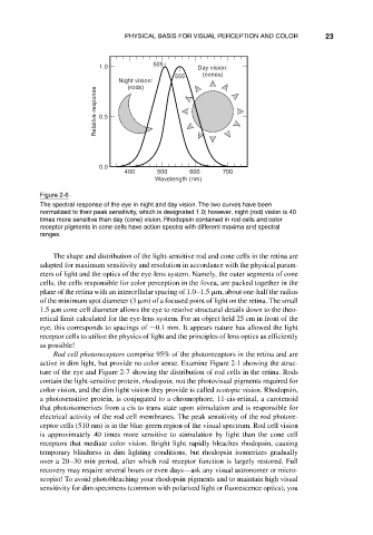
Figure 2-6
The spectral response of the eye in night and day vision. The two curves have been
normalized to their peak sensitivity, which is designated 1.0; however, night (rod) vision is 40
times more sensitive than day (cone) vision. Rhodopsin contained in rod cells and color
receptor pigments in cone cells have action spectra with different maxima and spectral
ranges.
The shape and distribution of the light-sensitive rod and cone cells in the retina are
adapted for maximum sensitivity and resolution in accordance with the physical param-
eters of light and the optics of the eye-lens system. Namely, the outer segments of cone
cells, the cells responsible for color perception in the fovea, are packed together in the
plane of the retina with an intercellular spacing of 1.0–1.5 m, about one-half the radius
of the minimum spot diameter (3 m) of a focused point of light on the retina. The small
1.5 m cone cell diameter allows the eye to resolve structural details down to the theo-
retical limit calculated for the eye-lens system. For an object held 25 cm in front of the
eye, this corresponds to spacings of 0.1 mm. It appears nature has allowed the light
receptor cells to utilize the physics of light and the principles of lens optics as efficiently
as possible!
Rod cell photoreceptors comprise 95% of the photoreceptors in the retina and are
active in dim light, but provide no color sense. Examine Figure 2-1 showing the struc-
ture of the eye and Figure 2-7 showing the distribution of rod cells in the retina. Rods
contain the light-sensitive protein, rhodopsin, not the photovisual pigments required for
color vision, and the dim light vision they provide is called scotopic vision. Rhodopsin,
a photosensitive protein, is conjugated to a chromophore, 11-cis-retinal, a carotenoid
that photoisomerizes from a cis to trans state upon stimulation and is responsible for
electrical activity of the rod cell membranes. The peak sensitivity of the rod photore-
ceptor cells (510 nm) is in the blue-green region of the visual spectrum. Rod cell vision
is approximately 40 times more sensitive to stimulation by light than the cone cell
receptors that mediate color vision. Bright light rapidly bleaches rhodopsin, causing
temporary blindness in dim lighting conditions, but rhodopsin isomerizes gradually
over a 20–30 min period, after which rod receptor function is largely restored. Full
recovery may require several hours or even days—ask any visual astronomer or micro-
scopist! To avoid photobleaching your rhodopsin pigments and to maintain high visual
sensitivity for dim specimens (common with polarized light or fluorescence optics), you