
Page 33 - Fundamentals of Light Microscopy and Electronic Imaging
P. 33
16 LIGHT AND COLOR
Cornea
Iris
Aqueous humor
Pupil
Lens
Retina
Blind spot Macula
and Fovea
Optic nerve
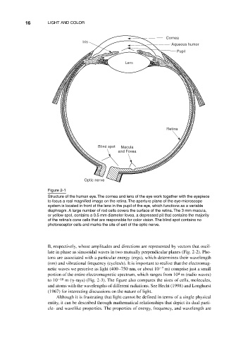
Figure 2-1
Structure of the human eye. The cornea and lens of the eye work together with the eyepiece
to focus a real magnified image on the retina. The aperture plane of the eye-microscope
system is located in front of the lens in the pupil of the eye, which functions as a variable
diaphragm. A large number of rod cells covers the surface of the retina. The 3 mm macula,
or yellow spot, contains a 0.5 mm diameter fovea, a depressed pit that contains the majority
of the retina’s cone cells that are responsible for color vision. The blind spot contains no
photoreceptor cells and marks the site of exit of the optic nerve.
B, respectively, whose amplitudes and directions are represented by vectors that oscil-
late in phase as sinusoidal waves in two mutually perpendicular planes (Fig. 2-2). Pho-
tons are associated with a particular energy (ergs), which determines their wavelength
(nm) and vibrational frequency (cycles/s). It is important to realize that the electromag-
netic waves we perceive as light (400–750 nm, or about 10 7 m) comprise just a small
4
portion of the entire electromagnetic spectrum, which ranges from 10 m (radio waves)
to 10 10 m ( -rays) (Fig. 2-3). The figure also compares the sizes of cells, molecules,
and atoms with the wavelengths of different radiations. See Hecht (1998) and Longhurst
(1967) for interesting discussions on the nature of light.
Although it is frustrating that light cannot be defined in terms of a single physical
entity, it can be described through mathematical relationships that depict its dual parti-
cle- and wavelike properties. The properties of energy, frequency, and wavelength are