
Page 215 - Handbook of Biomechatronics
P. 215
212 Georgios A. Bertos and Evangelos G. Papadopoulos
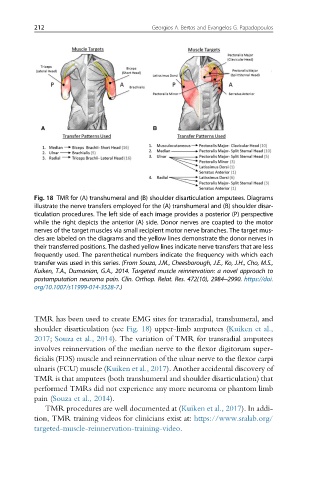
Fig. 18 TMR for (A) transhumeral and (B) shoulder disarticulation amputees. Diagrams
illustrate the nerve transfers employed for the (A) transhumeral and (B) shoulder disar-
ticulation procedures. The left side of each image provides a posterior (P) perspective
while the right depicts the anterior (A) side. Donor nerves are coapted to the motor
nerves of the target muscles via small recipient motor nerve branches. The target mus-
cles are labeled on the diagrams and the yellow lines demonstrate the donor nerves in
their transferred positions. The dashed yellow lines indicate nerve transfers that are less
frequently used. The parenthetical numbers indicate the frequency with which each
transfer was used in this series. (From Souza, J.M., Cheesborough, J.E., Ko, J.H., Cho, M.S.,
Kuiken, T.A., Dumanian, G.A., 2014. Targeted muscle reinnervation: a novel approach to
postamputation neuroma pain. Clin. Orthop. Relat. Res. 472(10), 2984–2990. https://doi.
org/10.1007/s11999-014-3528-7.)
TMR has been used to create EMG sites for transradial, transhumeral, and
shoulder disarticulation (see Fig. 18) upper-limb amputees (Kuiken et al.,
2017; Souza et al., 2014). The variation of TMR for transradial amputees
involves reinnervation of the median nerve to the flexor digitorum super-
ficialis (FDS) muscle and reinnervation of the ulnar nerve to the flexor carpi
ulnaris (FCU) muscle (Kuiken et al., 2017). Another accidental discovery of
TMR is that amputees (both transhumeral and shoulder disarticulation) that
performed TMRs did not experience any more neuroma or phantom limb
pain (Souza et al., 2014).
TMR procedures are well documented at (Kuiken et al., 2017). In addi-
tion, TMR training videos for clinicians exist at: https://www.sralab.org/
targeted-muscle-reinnervation-training-video.