Page 363 - Handbook of Biomechatronics
P. 363
Current Advances in the Design of Retinal and Cortical Visual Prostheses  357


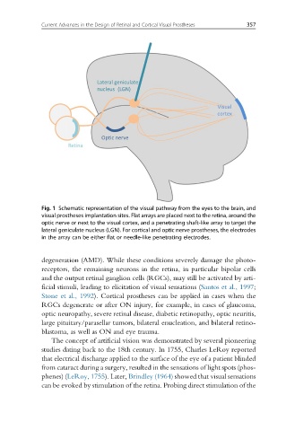
              Fig. 1 Schematic representation of the visual pathway from the eyes to the brain, and
              visual prostheses implantation sites. Flat arrays are placed next to the retina, around the
              optic nerve or next to the visual cortex, and a penetrating shaft-like array to target the
              lateral geniculate nucleus (LGN). For cortical and optic nerve prostheses, the electrodes
              in the array can be either flat or needle-like penetrating electrodes.


              degeneration (AMD). While these conditions severely damage the photo-
              receptors, the remaining neurons in the retina, in particular bipolar cells
              and the output retinal ganglion cells (RGCs), may still be activated by arti-
              ficial stimuli, leading to elicitation of visual sensations (Santos et al., 1997;
              Stone et al., 1992). Cortical prostheses can be applied in cases when the
              RGCs degenerate or after ON injury, for example, in cases of glaucoma,
              optic neuropathy, severe retinal disease, diabetic retinopathy, optic neuritis,
              large pituitary/parasellar tumors, bilateral enucleation, and bilateral retino-
              blastoma, as well as ON and eye trauma.
                 The concept of artificial vision was demonstrated by several pioneering
              studies dating back to the 18th century. In 1755, Charles LeRoy reported
              that electrical discharge applied to the surface of the eye of a patient blinded
              from cataract during a surgery, resulted in the sensations of light spots (phos-
              phenes) (LeRoy, 1755). Later, Brindley (1964) showed that visual sensations
              can be evoked by stimulation of the retina. Probing direct stimulation of the

