
Page 286 - Handbook of Deep Learning in Biomedical Engineering Techniques and Applications
P. 286
Chapter 10 Deep neural network in medical image processing 277
in the storage medium). Medical images are depicted by an array
of picture elements known as pixels or voxels, representing an
internal structure or feature of an anatomic region. This is a
discrete image resulting from a sampling/reconstruction process,
which maps numeric values into spatial positions. An indication
of the complexity in which the feature can be represented is the
number of pixels used to characterize the field-of-view of a
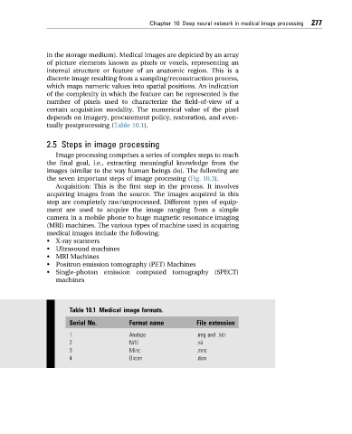
certain acquisition modality. The numerical value of the pixel
depends on imagery, procurement policy, restoration, and even-
tually postprocessing (Table 10.1).
2.5 Steps in image processing
Image processing comprises a series of complex steps to reach
the final goal, i.e., extracting meaningful knowledge from the
images (similar to the way human beings do). The following are
the seven important steps of image processing (Fig. 10.3).
Acquisition: This is the first step in the process. It involves
acquiring images from the source. The images acquired in this
step are completely raw/unprocessed. Different types of equip-
ment are used to acquire the image ranging from a simple
camera in a mobile phone to huge magnetic resonance imaging
(MRI) machines. The various types of machine used in acquiring
medical images include the following:
• X-ray scanners
• Ultrasound machines
• MRI Machines
• Positron emission tomography (PET) Machines
• Single-photon emission computed tomography (SPECT)
machines
Table 10.1 Medical image formats.
Serial No. Format name File extension
1 Analyze .img and .hdr
2 Nifti .nii
3 Minc .mnc
4 Dicom .dcm