
Page 43 - Handbook of Deep Learning in Biomedical Engineering Techniques and Applications
P. 43
Chapter 2 Deep convolutional neural network in medical image processing 31
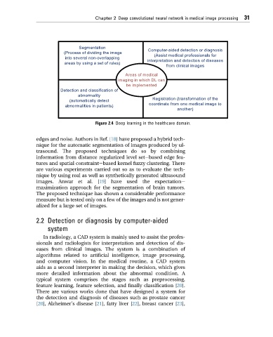
Segmentation
(Process of dividing the image Computer-aided detection or diagnosis
(Assist medical professionals for
into several non-overlapping
interpretation and detection of diseases
areas by using a set of rules)
from clinical images
Areas of medical
imaging in which DL can
be implemented
Detection and classification of
abnormality
Registration (transformation of the
(automatically detect
coordinate from one medical image to
abnormalities in patients)
another)
Figure 2.4 Deep learning in the healthcare domain.
edges and noise. Authors in Ref. [18] have proposed a hybrid tech-
nique for the automatic segmentation of images produced by ul-
trasound. The proposed techniques do so by combining
information from distance regularized level setebased edge fea-
tures and spatial constraintebased kernel fuzzy clustering. There
are various experiments carried out so as to evaluate the tech-
nique by using real as well as synthetically generated ultrasound
images. Anwar et al. [19] have used the expectatione
maximization approach for the segmentation of brain tumors.
The proposed technique has shown a considerable performance
measure but is tested only on a few of the images and is not gener-
alized for a large set of images.
2.2 Detection or diagnosis by computer-aided
system
In radiology, a CAD system is mainly used to assist the profes-
sionals and radiologists for interpretation and detection of dis-
eases from clinical images. The system is a combination of
algorithms related to artificial intelligence, image processing,
and computer vision. In the medical routine, a CAD system
aids as a second interpreter in making the decision, which gives
more detailed information about the abnormal condition. A
typical system comprises the stages such as preprocessing,
feature learning, feature selection, and finally classification [20].
There are various works done that have designed a system for
the detection and diagnosis of diseases such as prostate cancer
[20], Alzheimer's disease [21], fatty liver [22], breast cancer [23],