
Page 53 - Handbook of Deep Learning in Biomedical Engineering Techniques and Applications
P. 53
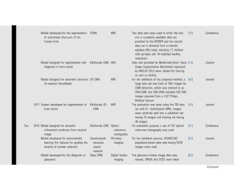
Conference       Journal   Journal       Journal     Conference  Journal  Conference  Continued


                    [78]             [79]      [80]          [50]        [81]   [82]      [83]
                                     Tumor     a  for  as  SWI
                    first     children  organized  training  to  data  set  having  optical  5378
                    the  set  second  rolandic  healthy  Brain  for  method,  images  320  Philips  TBI  images  validation  set  157  sets,  taken
                    which  data  the  a  17    proposed  SWI  referred  included  the  MRIs,  training  of  used  ACHIKO-NC  having  data  were

                    in   and  from  including  matched  Multimodal  Benchmark  utilized  of  was  3.0T  a  using  a  into  set  was  sets  image  SCES
                    used  available  BSIBSR  18  by  were  the  built  which  SWI-CMB  from  done  multichannel  split  and  a  process,  data
                    were  publicly  the  obtained  study,  and  Segmentation  2013  testing  of  was  set  the  System  was  images  purpose,  tomography  used  fundus  and
                    sets  a  by  is  set  (RE)  epilepsy  provided  MICCAI  as  validation  data  detection,  acquired  evaluation  61  randomly  15  validation  population-based  were  ORIGA
                    data  is  one  provided  data  epilepsy  with  individuals  sets  Image  well  the  large  CMB  SWI-CMB,  images  Medical  with  were  having  images  evaluation  coherence  the  images  glaucoma  namely,

                    Two              Data  by  as  For       The  set  46  For  For       The

                                                                           coherence  tomography  imaging  fundus  imaging


                    MRI              MRI       MRI           MRI         Optical  Slit-lamp  Digital

                                     CNN                     3D          CNN              CNN


                    FCNN             Multiscale  CNN  3D     Multiscale  CNN  Multiscale  Convolutional-  recursive  neural  network  Deep

                    segmentation  the  of  and  detection    of segmentation  medical  automatically  the  grading  of diagnosis


                    the  structures  segmentation  tumor  automatic      semantic  from  for  cataracts  the
                    for              for  brain  for  microbleeds  for   for  prediction  for  features  nuclear  for
                    developed  subcortical  brain  designed  of  designed  cerebral  developed  lesion  designed  information  developed  the  of  developed


                    Model  of  human  Model  diagnosis  Model  of  System  brain  Model  image  Model  learning  severity  Model  glaucoma

                                                             2017        2015


                                                                         Eye