
Page 24 - Handbook of Electronic Assistive Technology
P. 24
Chapter 1 • Basic Neurosciences With Relevance to Electronic Assistive Technology 11
excite and inhibit the peripheral motor nerves that in turn control the muscles involved in
the functional unit.
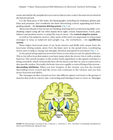
It is the deep parts of the brain, the basal ganglia, including the thalamus, globus pal-
lidus and putamen, that coordinate the basic descending control, upgrading and down-
grading signals – the locomotor driving system (Fig. 1-6).
Above and related to this we use our thinking, learning and communicating skills, coor-
dinating output using all our other inputs from sight, sound, temperature, touch, pain,
balance and position senses, to adapt the way we move – the cortical adaptive system.
As well as this adaptive section, other parts of the brain are important in using input
messages to keep us balanced and upright (e.g., the cerebellum) – the equilibrium
system.
These higher functional areas of our brain interact and fiddle with output from the
locomotor driving system, down from the brain stem to the spinal levels, coordinating
motor units to help us change our strength, direction and speed of movement (Fig. 1-7).
At the spinal and peripheral nerve level there occurs what we call the spinal reflex arc.
This is the reflex loop that makes your knee jump when the doctor hits it with a tendon
hammer. The stretch receptor in the tendon feeds signal back to the spinal cord that it’s
being stretched, which intrinsically fires off the motor unit that in turn is connected to
that specific tendon. Normally, the brain then acts rapidly to control firing of the system –
descending inhibition. When you lose integrity of the circuits, you lose the normal
descending motor control and the motor unit continues to fire (clonus) and you lose
motor function and ability.
The messages are then relayed out from the different spinal cord levels to the groups of
muscles that work in a motor unit, contracting and relaxing in turn to move us. Messages
FIGURE 1-6 Basal ganglia� Courtesy of Fig. 2-3 Barnes, L., Fairhurst, C., 2011. The Brain in Hemiplegia Handbook for
Parent and Professionals. Mackeith Press.