
Page 191 - Handbook of Properties of Textile and Technical Fibres
P. 191
168 Handbook of Properties of Textile and Technical Fibres
Table 5.5 Relative microstructure composition of Bombyx mori, a wild
silkworm, and spiders
Microstructure type Coil (%) a-Helices (%) b-Sheets (%) b-Turns (%)
B. mori 8 14 50 28
Samia cynthia ricini 10 14 45 31
Nephila clavipes 12 18 37 33
Nephila edulis 11 22 36 31
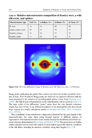
Figure 5.16 2D X-ray diffraction image of Bombyx mori silk fibers (l Co Ka ¼ 0.1789 nm).
Bragg peaks (indicating the partial fiber texture) are observed (in the crystalline struc-
ture at least, 30 to 50 narrow Bragg peaks are observed on a pattern collected with the
same procedure!); the notation of crystallographic planes comes from Drummy et al.
(2005). The Silk II unit-cell parameters of the orthorhombic cell are given in Table 5.5.
The large width of the diffraction “peaks” arises from the very limited coherence
length, less than 10 nm. X-ray diffraction patterns of the dried gland only show broad
rings, typical of a totally amorphous matter (Colomban and Dinh, 2012; Colomban
et al., 2012b).
The crystallinity of macromolecular compounds is hindered by the great length of the
macromolecules, the same chain going through regions of different degrees of
organization. Orientational disorder occurs readily for polymer backbones and hinders ac-
curate structure determination (Hosemann and Steffen, 1978). This point deserves further
studies and data must be read with caution, as most authors ignore the intrinsic variability
of the silk fibers, including its structure variability along their lengths (Fig. 5.5).