
Page 190 - Handbook of Properties of Textile and Technical Fibres
P. 190
Silk: fibers, films, and compositesdtypes, processing, structure, and mechanics 167
polyamide PA66 fiber with those of silks: different silkworms (B. mori, Gonometa
rufobrunea, Samia cynthia)or B. mori silk of different history: flotte, nondegummed
“gr ege” or degummed fibers, textile yarn, and regenerated films (Dinh et al., 2008).
DSC gives a global view of the polymer properties and structure. The trace of the
PA66 fiber shows a rather narrow endothermic (onset w250 C), characteristic of
the melting of a semicrystalline material (w50% of crystallinity (Marcellan et al.,
2004)) and a weak event related to the presence of water, at w120 C(<2%e3%
wt). In contrast, the melting peak of the different silks is three to four times broader,
related to a highly disordered/amorphous structure. A large mid-height width of the
peak is a maximum for regenerated silk. The shift of the onset of melting temperature
from w275 C(B. mori)to w360 C(S. cynthia) reveals the composition (amino
acids), macromolecule length, and structural differences between the fibroin chains.
Very small events are superimposed and can be associated with the crystallization
(if exothermic) or melting (endothermic) of minor phases. Desiccated glands appear
almost free of water although a strong water endothermic event is observed at
120e140 C for the fiber. Within B. mori silks, the highest melting temperature is
measured for the best quality of undegummed Chinese silk. A much broader melting
peak is measured for the regenerated film peaks due to its more amorphous character.
The crystallinity of regenerated films can, however, be preserved with optimized pro-
cedures. Different types of water are observed for regenerated films and for the silk
precursor according to their specific microstructures.
B. mori silk is generally described as being composed of a mixture of a “disordered”
Silk I (that found in the gland (Asakura et al., 2015a)) and a “crystallized” Silk II
(Askura et al., 2015a). The repetitive amino acid sequences GAGAGS are assumed
to promote intra- or extramolecular interaction leading to crystal creation called
b-sheet. Repetitive GAGAGY and separators lead to an amorphous or a-helix struc-
ture. Nevertheless, the polymer is not crystallized before spinning and is considered
to be, like Silk I, structured. Furthermore, models of transition of the crystalline part
from unspun Silk I to spun Silk II have been proposed from b-turns to b-sheets on
the basis of nuclear magnetic resonance techniques (Asakura et al., 1997, 2001,
2005, 2015a,b; Demura et al., 1998; Kameda et al., 2002), X-ray diffraction (Marsh
et al., 1955; Lotz and Cesari, 1979; Takahashi et al., 1999; R€ ossle, 2004; Jauzein,
2010; Colomban et al., 2012b), infrared spectroscopy (Chen et al., 2001; Taddei
and Monti, 2005; Hu et al., 2006a), circular dichroism (Wilson et al., 2000; Li
et al., 2001; Dicko et al., 2004; Termonia, 2004), and Raman scattering (Monti
et al., 2001; Rousseau et al., 2004; Taddei and Monti, 2005; Colomban et al.,
2008a, 2012a) analysis. Similar observations have been made for wild silkworms
(Nakazawa et al., 1999; Asakura and Nakazawa, 2004; Asakura et al., 2007; Magoshi
et al., 2000) and spiders (Parkhe et al., 1997; Lef evre et al., 2007a). There is consider-
able discussion in the literature as to the degree of crystallinity in different silks. Some
researchers have concluded that crystallinity is low (Colomban et al., 2008), whereas
others find much higher values, up to 50% (Lef evre et al., 2007b) for B. mori and from
w10% (Gosline et al., 1999) for spider silk in water to w30% for dry spider silk
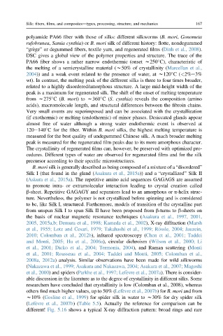
(Lef evre et al., 2007b)(Table 5.5). Actually the reference for comparison can be
different! Fig. 5.16 shows a typical X-ray diffraction pattern: broad rings and rare