
Page 324 - Industrial Ventilation Design Guidebook
P. 324
280 CHAPTER 5 PHYSIOLOGICAL AND TOXICOLOGICAL CONSIDERATIONS
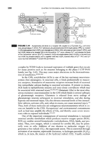
FIGURE 5.44 Acetylcholine (A) binds to a receptor (R) coupled to a G-protein (G p), and stimu-
lates phospholipase C (PLC). PLC hydrolyzes phosphatidylinositol-(4,5)-bisphosphate (PiP 2) to inositol-
(1,4,5)-trtphosphate (lns(l,4,5)P 3) and diacylglycerol (DAG). DAG stimulates protein kinase C (PKC),and
2+
2+
lns(l ,4,5)P3 binds to its receptor (R) in the intracellular Ca store, releases Ca , and elevates the levels
2
of free intracellular calcium {[Ca *],). lnositol-(l,3,4,5)-tetracisphosphate (lns(l,3,4,5)P 4) is formed from
2+
Ins(l,4,5)P 3 by phosphorylation, and it, together with lns(l,4,5)P 3 controls influx of Ca , PKC and Ca 2+
92
cause neuronal stimulation. (Used with permission.)
complex by TCDD leads to increased expression of multiple genes that encode
for many proteins such as the enzymes belonging to the phase I CYP P450
family (see Fig. 5.45). This may cause major alterations in the biotranstorma-
89 98 100
tion of xenobiotics. ' "
In the CNS, acetylcholine (ACh) is one of the key excitatory neurotrans-
mitters (first messengers). In neuronal cells, it binds preferentially to muscar-
ink receptors. Stimulation of muscarinic receptors increases levels of neuronal
free intracellular calcium leading to neuronal activation. If present in excess,
ACh leads to epileptiforrnic seizures and tonic-clonic convulsions which may
92 101 102
be associated with neuronal injury. ' ' Glutamate (Glu) is the most ubiq-
uitous excitatory neurotransmitter in the CNS which binds to several subtypes
of glutamatergic receptors. Glutamate is released from nerve endings of
glutamatergic neurons subsequent to neuronal stimulation, and also during
hypoxia and neuronal injury. Glu also elevates neuronal levels of free intracel-
93
lular calcium, activates cells, and, when in excess, can cause neuronal injury.
Thus, both of these molecules are endogenous neurotransmitters which in ex-
cess are harmful to the CNS. Occupational and environmental contaminants
such as lead may amplify the effects of Glu, and thereby cause severe neuro-
toxic risks to exposed individuals. 103 104
'
One of the important consequences of neuronal stimulation is increased
neuronal aerobic metabolism which produces reactive oxygen species (ROS).
ROS can oxidize several biomolecules (carbohydrates, DNA, lipids, and pro-
teins). Thus, even oxygen, which is essential for aerobic life, may be poten-
tially toxic to cells. Addition of one electron to molecular oxygen (O 2 )
generates a free radical (O^), the superoxide anion. This is converted through
activation of an enzyme, superoxide dismutase, to hydrogen peroxide (H 2O 2),
which is, in turn, the source of the hydroxyl radical (OH). Usually catalase