
Page 329 - Industrial Ventilation Design Guidebook
P. 329
5.3 TOXICiTY AND RISKS INDUCED BY OCCUPATIONAL EXPOSURE TO CHEMICAL COMPOUNDS 285
of cellular functions, and it always represents an unwanted effect on the cell
by a chemical. Apoptosis is a physiological phenomenon that is required dur-
ing development of the embryo in shaping the developing organs into their fi-
nal size and form, and it is also functionally important in the development of
organs and even body parts (e.g., fingers and toes). Apoptosis is also impor-
tant in maintaining the integrity and renewal of mucous membranes and the
skin. In direct contrast to necrosis which is a passive, non-energy-requiring
phenonomenon, apoptosis requires gene expression and synthesis of new pro-
89 91
teins, and it is an energy-expensive process. '
Necrotic cell death is often due to binding of reactive species to biologi-
cally important cellular macromolecules, such as proteins, lipids, and DNA.
Biotransformation of a number of chemicals such as carbon tetrachloride or
styrene leads to formation of epoxides that bind to nucleophilic sites on pro-
teins and DNA. Many of these compounds are also carcinogens. Furthermore,
several compounds also cause increased production of ROS. These phenom-
ena may also damage the cell membrane, leading to its leakage and rupture.
Necrosis is characterized by cell swelling and leakage of cell constituents into
the surroundings of the cell.
In apoptotic cell death, several factors such as growth factors, NO, the tu-
mor suppressor gene p53, and the protein encoded by this gene contribute to
the process that leads to cell death. One of the functions of p53 protein is the
activation of apoptosis if a cell is transformed to a malignant cell. Apoptosis
typically leads to the formation of smaller membrane-encapsulated particles
within the cell. Apoptotic cell death begins in the nucleus and proceeds to
other parts of the cell. The death process may be quite advanced before it can
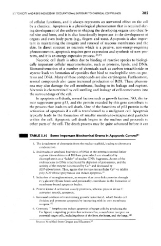
TABLE 5.10 Some Important Biochemical Events in Apoptotic Control 91
1. The detachment of chromatin from the nuclear scaffold, leading to chromatin
condensation.
2. Endonuclease-catalyzed hydrolysis of DNA at the internucleosomal linker
regions into multimers of 180 base pairs which are visualized by
electrophoresis as a "ladder" of nuclear DNA fragments. Access of the
endonuclease to DNA is facilitated by depletion of polyamines, and the
2+
activity of the enzyme is increased by Ca and decreased by
2+
ADP-ribosylation, Thus, agents that increase intracellular Ca or inhibit
poly(ADP-ribose) polymerase can induce apoptosis.' 21
3. Induction of transglutaminase, an enzyme that cross-links proteins through
£-(*y-glutamyl)iysine bonds and presumably contributes to the formation of
membrane-bound apoptosis bodies.
4. Protein kinase A activation usually promotes, whereas protein kinase C
activation retards, apoptosis.
5. Increased synthesis of transforming growth factor-betal, which blocks cell
division and promotes apoptosis by interacting with its own membrane
receptor. 122
6. Cytotoxic T lymphocytes induce apoptosis of target cells by producing the
Fas ligand, a signaling protein that activates Fas, a membrane receptor on
potential target cells, including those of the liver, the heart, and the lungs. 123
Source: Modified from Gregus and Klaassen. 89