
Page 261 - Inorganic Mass Spectrometry - Fundamentals and Applications
P. 261
247
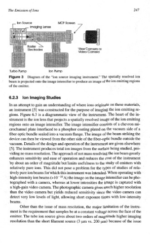
Diagram of the “ion source imaging ins~ment.” The spatially resolved ion
beam is projected onto the image intensifier to produce an image of the ion emitting regions
of the emitter.
In an attempt to gain an understanding of where ions originate on these materials,
an instrument [53 was constructed for the purpose of imaging the ion emitting re-
gions. Figure 6.3 is a diagrammatic view of the instrument. The heart of the in-
of
strument is the ion lens that projects a spatially resolved image the ion emitting
regions onto an image intensifier. The image intensifier consists of a chevron mi-
crochannel plate interfaced to a phosphor coating plated on the vacuum side
a
of
fiber optic bundle sealed into a vacuum flange. The image of the beam striking the
device can then be viewed from the other side of the fiber-optic bundle outside the
vacuum. Details of the design and operation of the instrument are given elsewhere
[5]. The instrument produces total ion images from the surface being studied, pro-
viding no mass resolution. The approach of not mass resolving the ion beam greatly
enhances sensitivity and ease of operation and reduces the cost of the instru~ent
by about an order of magnitude but lirnits usefulness the study of emitters with
to
relatively pure ions. This did not pose a problem for the types of studies of rela-
tively pure ion beams forwhich this instrument was intended. When operating with
high-intensity ion beams A) the image on the image intensifier can be pho-
tographed with a camera, whereas at lower intensities the image is captured with
a high-gain video camera. The photographic camera gives much higher resolution
than the video cmera but yields reduced sensitivity since the video camera can
detect very low levels of light, allowing short exposure times with low-intensity
~ beams.
Other than the issue of mass resolution, the major limitation of the instru-
ment is the requirement that samples be at a constant voltage across the face of the
emitter. The tube ion source gives about two orders magnitude higher imaging
of
resolution than the short filament source (3 pm vs. 200 pm) because of the issue