
Page 157 - Microtectonics
P. 157
146 5 · Shear Zones
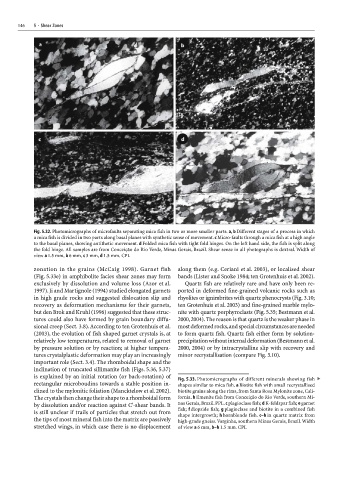
Fig. 5.32. Photomicrographs of microfaults separating mica fish in two or more smaller parts. a, b Different stages of a process in which
a mica fish is divided in two parts along basal planes with synthetic sense of movement. c Micro-faults through a mica fish at a high angle
to the basal planes, showing antithetic movement. d Folded mica fish with tight fold hinges. On the left hand side, the fish is split along
the fold hinge. All samples are from Conceição do Rio Verde, Minas Gerais, Brazil. Shear sense in all photographs is dextral. Width of
view a 1.5 mm, b 6 mm, c 3 mm, d 1.5 mm. CPL
zonation in the grains (McCaig 1998). Garnet fish along them (e.g. Ceriani et al. 2003), or localised shear
(Fig. 5.33e) in amphibolite facies shear zones may form bands (Lister and Snoke 1984; ten Grotenhuis et al. 2002).
exclusively by dissolution and volume loss (Azor et al. Quartz fish are relatively rare and have only been re-
1997). Ji and Martignole (1994) studied elongated garnets ported in deformed fine-grained volcanic rocks such as
in high grade rocks and suggested dislocation slip and rhyolites or ignimbrites with quartz phenocrysts (Fig. 3.10;
recovery as deformation mechanisms for their garnets, ten Grotenhuis et al. 2003) and fine-grained marble mylo-
but den Brok and Kruhl (1996) suggested that these struc- nite with quartz porphyroclasts (Fig. 5.35; Bestmann et al.
tures could also have formed by grain boundary diffu- 2000, 2004). The reason is that quartz is the weaker phase in
sional creep (Sect. 3.8). According to ten Grotenhuis et al. most deformed rocks, and special circumstances are needed
(2003), the evolution of fish shaped garnet crystals is, at to form quartz fish. Quartz fish either form by solution-
relatively low temperatures, related to removal of garnet precipitation without internal deformation (Bestmann et al.
by pressure solution or by reaction; at higher tempera- 2000, 2004) or by intracrystalline slip with recovery and
tures crystalplastic deformation may play an increasingly minor recrystallisation (compare Fig. 3.10).
important role (Sect. 3.4). The rhomboidal shape and the
inclination of truncated sillimanite fish (Figs. 5.36, 5.37)
is explained by an initial rotation (or back-rotation) of
Fig. 5.33. Photomicrographs of different minerals showing fish ▼
rectangular microboudins towards a stable position in- shapes similar to mica fish. a Biotite fish with small recrystallised
clined to the mylonitic foliation (Mancktelow et al. 2002). biotite grains along the rims, from Santa Rosa Mylonite zone, Cali-
The crystals then change their shape to a rhomboidal form fornia. b Ilmenite fish from Conceição do Rio Verde, southern Mi-
by dissolution and/or reaction against C'-shear bands. It nas Gerais, Brazil. PPL. c plagioclase fish; d K-feldspar fish; e garnet
is still unclear if trails of particles that stretch out from fish; f diopside fish; g plagioclase and biotite in a combined fish
shape intergrowth; h hornblende fish. c–h in quartz matrix from
the tips of most mineral fish into the matrix are passively high-grade gneiss. Varginha, southern Minas Gerais, Brazil. Width
stretched wings, in which case there is no displacement of view a 6 mm, b–h 1.5 mm. CPL