
Page 154 - Microtectonics
P. 154
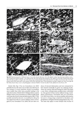
5.6  ·  Microscopic Shear Sense Indicators in Mylonite  143


                 Fig. 5.29. Photomicrographs of different types of mica fish. a Lenticular mica fish with slightly inclined tips showing undulose extinc-
                 tion, transitional between groups 1 and 2; b lenticular fish with internal discontinuity in the right tip of the fish; c rhomboidal shaped
                 fish with (001) parallel to longest side of the fish, group 3; d rhomboidal shaped fish with (001) parallel to the shortest side of the fish,
                 group 4; e fish with small aspect ratio, group 5; f mica fish with high aspect ratio belonging to group 6. Samples are from Conceição do
                 Rio Verde, Brazil. Shear sense in all photographs is dextral. Width of view a 3 mm, b 0.75 mm, c, d and e 3 mm, f 6 mm. CPL
                   Biotite fish (Fig. 5.33a; ten Grotenhuis et al. 2003)  dence of internal deformation and were interpreted to
                 probably form in the same way as white mica fish but are  have formed essentially by grain size reducing mecha-
                 less common. In most mylonites, biotite recrystallises  nisms. By contrast, Ishii and Sawaguchi (2002) described
                 more readily than white mica, possibly leading to the  a case where orthopyroxene porphyroclasts embedded
                 early destruction of biotite fish. K-feldspar fish in a quartz  in a fine-grained olivine matrix suffered strong crystal-
                 feldspar matrix (ten Grotenhuis et al. 2003) in high-grade  plastic deformation. Tourmaline fish (ten Grotenhuis
                 rocks may also form by internal deformation (Fig. 5.33d),  et al. 2003) show little or no intracrystalline deformation
                 possibly assisted by dynamic recrystallisation along the  but seem to change shape exclusively by dissolution and
                 rim. Hyperstene fish in a quartz-feldspar matrix investi-  precipitation, cutting the internal zoning patterns (Fig. 5.34).
                 gated by ten Grotenhuis et al. (2003) did not show evi-  The same may apply to some feldspar fish, because of