
Page 153 - Microtectonics
P. 153
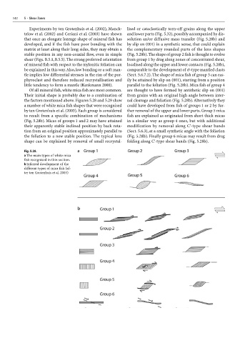
142   5  ·  Shear Zones
                     Experiments by ten Grotenhuis et al. (2002), Manck-  lised or cataclastically torn-off grains along the upper
                   telow et al. (2002) and Ceriani et al. (2003) have shown  and lower parts (Fig. 5.32), possibly accompanied by dis-
                   that once an elongate lozenge shape of mineral fish has  solution an/or diffusive mass transfer (Fig. 5.28b) and
                   developed, and if the fish have poor bonding with the  by slip on (001) in a synthetic sense, that could explain
                   matrix at least along their long sides, they may obtain a  the complementary rounded parts of the lens shapes
                   stable position in any non-coaxial flow, even in simple  (Fig. 5.28b). The shape of group 2 fish is thought to evolve
                   shear (Figs. B.5.1, B.5.5). The strong preferred orientation  from group 1 by drag along zones of concentrated shear,
                   of mineral fish with respect to the mylonitic foliation can  localised along the upper and lower contacts (Fig. 5.28b),
                   be explained in this way. Also, low bonding or a soft man-  comparable to the development of σ-type mantled clasts
                   tle implies low differential stresses in the rim of the por-  (Sect. 5.6.7.2). The shape of mica fish of group 3 can eas-
                   phyroclast and therefore reduced recrystallisation and  ily be attained by slip on (001), starting from a position
                   little tendency to form a mantle (Kenkmann 2000).  parallel to the foliation (Fig. 5.28b). Mica fish of group 4
                     Of all mineral fish, white mica fish are most common.  are thought to have formed by antithetic slip on (001)
                   Their initial shape is probably due to a combination of  from grains with an original high angle between inter-
                   the factors mentioned above. Figures 5.28 and 5.29 show  nal cleavage and foliation (Fig. 5.28b). Alternatively they
                   a number of white mica fish shapes that were recognised  could have developed from fish of groups 1 or 2 by fur-
                   by ten Grotenhuis et al. (2003). Each group is considered  ther removal of the upper and lower parts. Group 5 mica
                   to result from a specific combination of mechanisms  fish are explained as originated from short thick micas
                   (Fig. 5.28b). Micas of groups 1 and 2 may have attained  in a similar way as group 4 ones, but with additional
                   their apparently stable inclined position by back rota-  modification by removal along C'-type shear bands
                   tion from an original position approximately parallel to  (Sect. 5.6.3), at a small synthetic angle with the foliation
                   the foliation to a new stable position. The typical lens  (Fig. 5.28b). Finally group 6 micas may result from drag
                   shape can be explained by removal of small recrystal-  folding along C'-type shear bands (Fig. 5.28b).

                   Fig. 5.28.
                   a The main types of white mica
                   fish recognised in thin section.
                   b Inferred development of the
                   different types of mica fish (af-
                   ter ten Grotenhuis et al. 2003)