
Page 95 - MODERN ELECTROCHEMISTRY
P. 95
38 CHAPTER 2
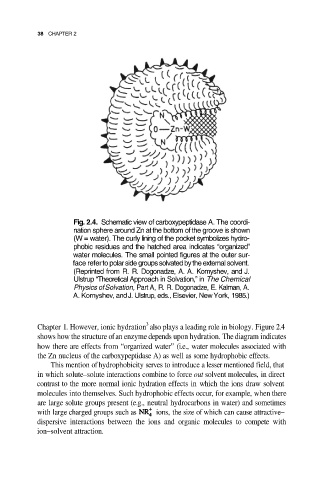
Fig. 2.4. Schematic view of carboxypeptidase A. The coordi-
nation sphere around Zn at the bottom of the groove is shown
(W = water). The curly lining of the pocket symbolizes hydro-
phobic residues and the hatched area indicates “organized”
water molecules. The small pointed figures at the outer sur-
face refer to polar side groups solvated by the external solvent.
(Reprinted from R. R. Dogonadze, A. A. Komyshev, and J.
Ulstrup “Theoretical Approach in Solvation,” in The Chemical
Physics of Solvation, Part A, R. R. Dogonadze, E. Kalman, A.
A. Kornyshev, and J. Ulstrup, eds., Elsevier, New York, 1985.)
3
Chapter 1. However, ionic hydration also plays a leading role in biology. Figure 2.4
shows how the structure of an enzyme depends upon hydration. The diagram indicates
how there are effects from “organized water” (i.e., water molecules associated with
the Zn nucleus of the carboxypeptidase A) as well as some hydrophobic effects.
This mention of hydrophobicity serves to introduce a lesser mentioned field, that
in which solute–solute interactions combine to force out solvent molecules, in direct
contrast to the more normal ionic hydration effects in which the ions draw solvent
molecules into themselves. Such hydrophobic effects occur, for example, when there
are large solute groups present (e.g., neutral hydrocarbons in water) and sometimes
with large charged groups such as ions, the size of which can cause attractive–
dispersive interactions between the ions and organic molecules to compete with
ion–solvent attraction.