
Page 176 - Modern Optical Engineering The Design of Optical Systems
P. 176
Characteristics of the Human Eye 159
fact, a reasonable simulation of the optics of the eye can be made by
considering the eye as a single refracting surface of water
(n D 1.333, V 55).
The following table lists typical values for the radii, thicknesses, and
indices of the optical surfaces of the eye. These, of course, vary from
individual to individual.
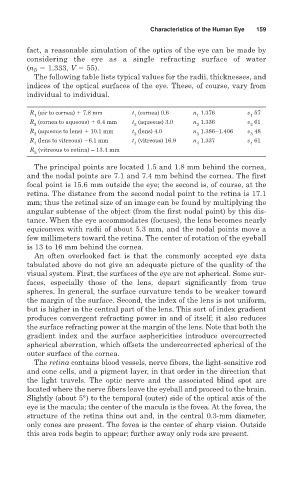
R (air to cornea) 7.8 mm t (cornea) 0.6 n 1.376 v 57
1 1 1 1
R (cornea to aqueous) 6.4 mm t (aqueous) 3.0 n 1.336 v 61
2 2 2 2
R (aqueous to lens) 10.1 mm t (lens) 4.0 n 1.386–1.406 v 48
3 3 3 3
R (lens to vitreous) 6.1 mm t (vitreous) 16.9 n 1.337 v 61
4 4 4 4
R (vitreous to retina) – 13.4 mm
5
The principal points are located 1.5 and 1.8 mm behind the cornea,
and the nodal points are 7.1 and 7.4 mm behind the cornea. The first
focal point is 15.6 mm outside the eye; the second is, of course, at the
retina. The distance from the second nodal point to the retina is 17.1
mm; thus the retinal size of an image can be found by multiplying the
angular subtense of the object (from the first nodal point) by this dis-
tance. When the eye accommodates (focuses), the lens becomes nearly
equiconvex with radii of about 5.3 mm, and the nodal points move a
few millimeters toward the retina. The center of rotation of the eyeball
is 13 to 16 mm behind the cornea.
An often overlooked fact is that the commonly accepted eye data
tabulated above do not give an adequate picture of the quality of the
visual system. First, the surfaces of the eye are not spherical. Some sur-
faces, especially those of the lens, depart significantly from true
spheres. In general, the surface curvature tends to be weaker toward
the margin of the surface. Second, the index of the lens is not uniform,
but is higher in the central part of the lens. This sort of index gradient
produces convergent refracting power in and of itself; it also reduces
the surface refracting power at the margin of the lens. Note that both the
gradient index and the surface asphericities introduce overcorrected
spherical aberration, which offsets the undercorrected spherical of the
outer surface of the cornea.
The retina contains blood vessels, nerve fibers, the light-sensitive rod
and cone cells, and a pigment layer, in that order in the direction that
the light travels. The optic nerve and the associated blind spot are
located where the nerve fibers leave the eyeball and proceed to the brain.
Slightly (about 5°) to the temporal (outer) side of the optical axis of the
eye is the macula; the center of the macula is the fovea. At the fovea, the
structure of the retina thins out and, in the central 0.3-mm diameter,
only cones are present. The fovea is the center of sharp vision. Outside
this area rods begin to appear; further away only rods are present.