
Page 127 - Nanotechnology an introduction
P. 127
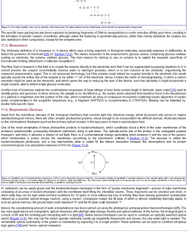
Figure 11.3 Four oligonucleotides, which can only assemble in the manner shown. The dashes represent strong covalent bonds, and the dashed lines represent weak hydrogen bonds.
  The specific base-pairing has also been exploited by fastening fragments of DNA to nanoparticles to confer selective affinity upon them, resulting in
  the formation of specific clusters of particles, although unless the fastening is geometrically precise, rather than merely statistical, the clusters are
  also statistical in their composition, except for the simplest ones.
  11.5. Biosensors
  The dictionary definition of a biosensor is “a device which uses a living organism or biological molecules, especially enzymes or antibodies, to
  detect the presence of chemicals”[35] (cf. Section 7.9.2). The classic biosensor is the amperometric glucose sensor, comprising glucose oxidase
  coating  an  electrode;  the  enzyme  oxidizes  glucose.  The  main  reason  for  wishing  to  use  an  enzyme  is  to  exploit  the  exquisite  specificity  of
  biomolecular binding interactions (“molecular recognition”).

  The Holy Grail of research in the field is to couple the enzyme directly to the electrode such that it can be regenerated by passing electrons to it; in
  current  practice  the  enzyme  concomitantly  reduces  water  to  hydrogen  peroxide,  which  is  in  turn  reduced  at  the  electrode,  engendering  the
  measured amperometric signal. This is not nanoscale technology, but if the enzyme could indeed be coupled directly to the electrode, this would
  typically require the active site of the enzyme to be within ~1 nm of the electrode, hence it enters the realm of nanoengineering, in which a carbon
  nanotube might be used as the electrode, and which opens the way to reducing the size of the device, such that ultimately it might incorporate a
  single enzyme, able to detect single glucose molecules.
  Another kind of biosensor exploits the combinatorial uniqueness of base strings of even fairly modest length to fabricate “gene chips”[33] used to
  identify genes and genomes. In these devices, the sample to be identified (e.g., the nucleic acids extracted from bacteria found in the bloodstream
  of a patient) is dispersed over the surface of the chip, which comprises an array of contiguous microzones containing known oligomers of nucleic
  acids complementary to the sought-for sequences (e.g., a fragment GATTACA is complementary to CTAATGA). Binding can be detected by
  double helix-specific dyes.
  11.6. Biophotonic Devices
  Apart from the marvellous intricacy of the biological machinery that converts light into chemical energy, which at present only serves to inspire
  nanotechnological mimics, there are other, simpler, photoactive proteins, robust enough to be incorporated into artificial devices. Molecules based
  on the chromophore rhodopsin (such as the primary optical receptor in the eye) seem to have a special place here.

  One of the most remarkable of these photoactive proteins is bacteriorhodopsin, which constitutes about a third of the outer membranes of the
  archaeon (extremophilic prokaryote) Halobium salinarum, living in salt lakes. The optically active site of the protein is the conjugated polyene
  rhodopsin, and when it absorbs a photon of red light, there is a conformational change generating strain between it and the rest of the protein,
  which  translocates  a  proton  across  the  membrane  (according  to  the  mechanism  outlined  in Section  11.3).  The  process  is  called  the
  bacteriorhodopsin  photocycle,  and  a  key  intermediate  state  is  called  M;  the  altered  interaction  between  the  chromophore  and  its  protein
  environment gives it an absorption maximum of 410 nm (Figure 11.4).


  Figure 11.4 Simplified view of the bacteriorhodopsin photocycle. A 570 nm photon absorbed by the ground state bR 570  (the subscript indicates the wavelength of maximum adsorption of the molecule) rapidly (within a few microseconds)
  transforms (through a series of intermediate stages) the molecule to the relatively stable intermediate M 410 . This state slowly relaxes thermally back to the ground state, but it can also be rapidly converted by a 410 nm photon. The
  thermal stability of the M state can be extended almost indefinitely by genetically modifying the protein.
  H. salinarum can be easily grown and the bacteriorhodopsin harvested in the form of “purple membrane fragments”—pieces of outer membrane
  consisting of an array of bacteriorhodopsin with the membrane lipid filling the interstitial volume. These fragments can be oriented and dried, in
  which state they can be kept under ambient conditions for 10 years or more without any loss of activity; they have already generated considerable
  interest as a possible optical storage medium, using a bacteri- orhodopsin mutant, the M state of which is almost indefinitely thermally stable. In
  such an optical memory, the ground state could represent “0” and the M state could represent “1”.
  Indeed, the overwhelming amount of work in biophotonics has been carried out using the photoactive archaeal protein bacteriorhodopsin (bR). The
  two main applications are holographic optical memories with ultrahigh data storage density and optical switches. In the former, the biological part is
  a block of bR and the nonliving part interacting with it is light [68]. Native bacteriorhodopsin can be used to construct an optically switched optical
  switch (Figure 11.5). Not only can the switch operate extremely rapidly (at megahertz frequencies and above), but only weak light is needed. The
  remarkable optical nonlinearity of the protein is manifested by exposing it to a single photon! These switches can be used to construct all-optical
  logic gates [168] and, hence, optical computers.