
Page 483 - New Trends in Eco efficient and Recycled Concrete
P. 483
Microstructural studies on recycled aggregate concrete 433
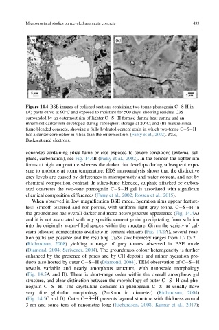
Figure 14.4 BSE images of polished sections containing two-tonne phenograin C S-H in:
(A) paste cured at 90 C and exposed to moisture for 500 days, showing residual C3S
surrounded by an outermost rim of lighter C S H formed during heat curing and an
innermost darker rim developed during subsequent storage at 20 C; and (B) mature silica
fume blended concrete, showing a fully hydrated cement grain in which two-tonne C S H
has a darker core richer in silica than the outermost rim (Famy et al., 2002). BSE,
Backscattered electrons.
concretes containing silica fume or else exposed to severe conditions (external sul-
phate, carbonation), see Fig. 14.4B(Famy et al., 2002). In the former, the lighter rim
forms at high temperature whereas the darker rim develops during subsequent expo-
sure to moisture at room temperature; EDS microanalysis shows that the distinctive
grey levels are caused by differences in microporosity and water content, and not by
chemical composition contrast. In silica-fume blended, sulphate attacked or carbon-
ated concretes the two-tone phenograin C S H gel is associated with significant
chemical composition differences (Famy et al., 2002; Rossen et al., 2015).
When observed in low magnification BSE mode, hydration rims appear feature-
less, smooth-textured and non-porous, with uniform light grey tonne. C S Hin
the groundmass has overall darker and more heterogeneous appearance (Fig. 14.4A)
and it is not associated with any specific cement grain, precipitating from solution
into the originally water-filled spaces within the structure. Given the variety of cal-
cium silicates compositions available in cement clinkers (Fig. 14.2A), several reac-
tion paths are possible and the resulting Ca/Si stoichiometry ranges from 1.2 to 2.1
(Richardson, 2008) yielding a range of grey tonnes observed in BSE mode
(Diamond, 2004; Scrivener, 2004). The groundmass colour heterogeneity is further
enhanced by the presence of pores and by CH deposits and minor hydration pro-
ducts also hosted by outer C S H(Diamond, 2004). TEM observation of C S H
reveals variable and nearly amorphous structure, with nanoscale morphology
(Fig. 14.5A and B). There is short-range order within the overall amorphous gel
structure, and clear distinction between the morphology of outer C S H and phe-
nograin C S H. The crystalline domains in phenograin C S H usually have
very fine globular morphology (2 8 nm in diameter) (Richardson, 2004)
(Fig. 14.5C and D). Outer C S H presents layered structure with thickness around
3 nm and some tens of nanometre long (Richardson, 2008; Kumar et al., 2017);