
Page 103 - Optofluidics Fundamentals, Devices, and Applications
P. 103
84 Cha pte r F i v e
polystyrene beads as small as 350 nm in diameter using interfering
Gaussian beams reflected off a prism surface. Grigorenko et al. [43]
also recently extended earlier approaches to surface plasmon reso-
nance (SPR)-based optical manipulation by exploiting the localized
plasmonic resonance in surface-bound metallic nanostructures. While
in general these methods are successful at trapping and even assem-
bling [44] small particles, they are limited by the distance through
which they can transport objects, since the optical manipulation
region is limited by the field of view of the focused laser, and the
plasmon propagation distance is relatively short.
The first clear demonstrations of long-distance optical transport
on waveguides focused on the use of solid-core, fluid-clad structures
that relied on the evanescent field of the waveguide to both capture
and transport suspended particles. These experiments featured the
propulsion of a wide variety of materials, organic and inorganic, on
waveguides. Kawata and Sugiura [50], for example, first demonstrated
the use of an evanescent field-based optical trapping technique. This
was further refined in 2000 by Tanaka and Yamamoto [51], who showed
the propulsion of polystyrene spheres on a channel waveguide.
While these seminal papers demonstrated for the first time the
potential for using evanescent field trapping as a potential mecha-
nism for optofluidic transport, it was unknown if the method would
have the same versatility demonstrated for optical tweezers. Gaugi-
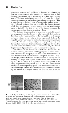
ran et al. [52] demonstrated the use of silicon nitride waveguides for
trapping and propulsion of yeast and red blood cells, as shown in
Fig. 5-3. The advantage in using silicon nitride waveguides is the
ability to guide wavelengths of light at 1064 nm. Unlike silicon
waveguides, which optimally guide light at telecom frequencies, at
1064 nm the light is not heavily absorbed by water, therefore reduc-
ing the impact on biological species. In addition, with a smaller
Light
→
F 10 μm 10 μm
(a) (b) (c)
FIGURE 5-3 Optofl uidic transport of biological species. (a) Finite element simulation
of optical fi eld in a channel waveguide and forces acting on a glass particle.
(b) Image of radiation pressure transport of red blood cells. (c) Yeast cells.
(S. Gaugiran, S. Getin, J. M. Fedeli, G. Colas, A. Fuchs, F. Chatelain, and J. Derouard,
“Optical manipulation of microparticles and cells on silicon nitride waveguides,” Optics
Express, 13(18), (2005), 6956–6963.) (See also color insert.)