
Page 172 - Principles of Catalyst Development
P. 172
160 CHAPTER 7
where B is the peak width for a diffraction line at angle 0, b is the value
for a well crystallized specimen, and A is the wavelength.(m)
Reliable data are only possible down to 5 nm since smaller crystallites
give such broad lines that sensitivity decreases. An advantage to this method
is that the sample may be heated and exposed to reactive atmospheres, and
changes in size during dynamic conditions are observable. Appropriate line
profile analysis measures crystallite size distributionsY24) When perfected,
this approach will be a valuable adjunct to morphology characterization.
Small angle x-ray scattering (SAXS) has been used in the past to study
pore size distributions in amorphous materials. The method gives good
results but is not now widely practiced.(22])
7.4.1.4. Extended X-Ray Absorption Fine Structure (EXAFS)
This is a relatively new tool that shows great promise. X-rays, when
absorbed, transmit photon energy to inner electrons, which then escape
from the atom. Interaction between these electrons and neighboring atoms
produce fine structure in the x-ray absorption edge, giving information on
coordination numbers and interatomic distance. Unfortunately, high
intensity x-rays from synchrotons are necessary, so that the technique is
not readily available. Nevertheless, valuable information on surface environ-
ments, not available from other sources, is beginning to appear.(II)
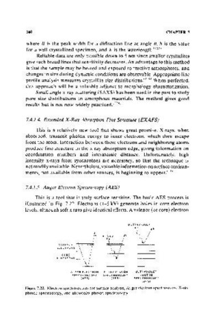
7.4.1.5. Auger Electron Spectroscopy (AES)
This is a tool that is truly surface sensitive. The basic AES process is
illustrated in Fig. 7.22. Electrons (1-5 kV) generate holes in core electron
levels, although soft x rays give identical effects. A valence (or core) electron
ULTRAVIOLET
/e X-hrJ'f' r "~
E~~~~~~~s ~e
,
CORE
ELECTRONS
AUGER ELECTRON X-RAY PHOTON ULTRAVIOLET
SPECTROSCOPY SPECTROSCOPY PHOTON
(AES) (XPS) SPECTROSCOPY
(UPS)
Figure 7.22. Electron spectroscopies for surface analysis, Auger electron spectroscopy, X-ray
photon spectroscopy, and ultraviolet photon spectroscopy.