
Page 63 - The Biochemistry of Inorganic Polyphosphates
P. 63
March 9, 2004
Char Count= 0
15:30
WU095-04
WU095/Kulaev
Complexes of polyphosphates with nucleic acids 47
−
−
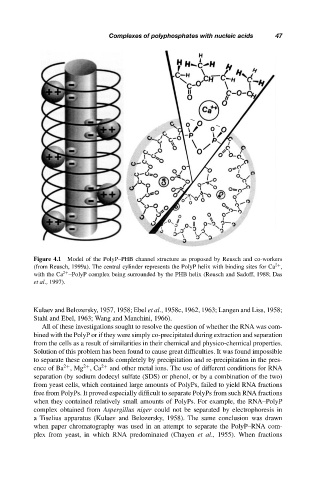
Figure 4.1 Model of the PolyP–PHB channel structure as proposed by Reusch and co-workers
(from Reusch, 1999a). The central cylinder represents the PolyP helix with binding sites for Ca ,
2+
with the Ca –PolyP complex being surrounded by the PHB helix (Reusch and Sadoff, 1988; Das
2+
et al., 1997).
Kulaev and Belozersky, 1957, 1958; Ebel et al., 1958c, 1962, 1963; Langen and Liss, 1958;
Stahl and Ebel, 1963; Wang and Manchini, 1966).
All of these investigations sought to resolve the question of whether the RNA was com-
bined with the PolyP or if they were simply co-precipitated during extraction and separation
from the cells as a result of similarities in their chemical and physico-chemical properties.
Solution of this problem has been found to cause great difficulties. It was found impossible
to separate these compounds completely by precipitation and re-precipitation in the pres-
2+
2+
ence of Ba ,Mg ,Ca 2+ and other metal ions. The use of different conditions for RNA
separation (by sodium dodecyl sulfate (SDS) or phenol, or by a combination of the two)
from yeast cells, which contained large amounts of PolyPs, failed to yield RNA fractions
free from PolyPs. It proved especially difficult to separate PolyPs from such RNA fractions
when they contained relatively small amounts of PolyPs. For example, the RNA–PolyP
complex obtained from Aspergillus niger could not be separated by electrophoresis in
a Tiselius apparatus (Kulaev and Belozersky, 1958). The same conclusion was drawn
when paper chromatography was used in an attempt to separate the PolyP–RNA com-
plex from yeast, in which RNA predominated (Chayen et al., 1955). When fractions