
Page 64 - The Biochemistry of Inorganic Polyphosphates
P. 64
WU095/Kulaev
WU095-04
Forms of polyphosphates cells
48 March 9, 2004 15:30 Char Count= 0
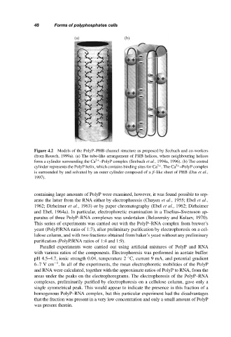
(a) (b)
Figure 4.2 Models of the PolyP–PHB channel structure as proposed by Seebach and co-workers
(from Reusch, 1999a). (a) The tube-like arrangement of PHB helices, where neighbouring helices
form a cylinder surrounding the Ca –PolyP complex (Seebach et al., 1994a, 1996). (b) The central
2+
cylinder represents the PolyP helix, which contains binding sites for Ca . The Ca –PolyP complex
2+
2+
is surrounded by and solvated by an outer cylinder composed of a β-like sheet of PHB (Das et al.,
1997).
containing large amounts of PolyP were examined, however, it was found possible to sep-
arate the latter from the RNA either by electrophoresis (Chayen et al., 1955; Ebel et al.,
1962; Dirheimer et al., 1963) or by paper chromatography (Ebel et al., 1962; Dirheimer
and Ebel, 1964a). In particular, electrophoretic examination in a Tiselius–Swensson ap-
paratus of three PolyP–RNA complexes was undertaken (Belozersky and Kulaev, 1970).
This series of experiments was carried out with the PolyP–RNA complex from brewer’s
yeast (PolyP/RNA ratio of 1:7), after preliminary purification by electrophoresis on a cel-
lulose column, and with two fractions obtained from baker’s yeast without any preliminary
purification (PolyP/RNA ratios of 1:4 and 1:9).
Parallel experiments were carried out using artificial mixtures of PolyP and RNA
with various ratios of the components. Electrophoresis was performed in acetate buffer:
pH 4.5–4.7, ionic strength 0.04, temperature 2 C, current 9 mA, and potential gradient
◦
−1
6–7Vcm . In all of the experiments, the mean electrophoretic mobilities of the PolyP
and RNA were calculated, together with the approximate ratios of PolyP to RNA, from the
areas under the peaks on the electrophoregrams. The electrophoresis of the PolyP–RNA
complexes, preliminarily purified by electrophoresis on a cellulose column, gave only a
single symmetrical peak. This would appear to indicate the presence in this fraction of a
homogenous PolyP–RNA complex, but this particular experiment had the disadvantages
that the fraction was present in a very low concentration and only a small amount of PolyP
was present therein.