
Page 151 - Vibrational Spectroscopic Imaging for Biomedical Applications
P. 151
sFTIR, Raman, and SERS Imaging of Fungal Cells 127
dilemma, that of the biology and the spectroscopy being equally
attractive avenues to pursue; we continue to travel down both
roads.
5.2 Introduction to Fungi
Fungi are eukaryotic microbes whose complex internal cellular
architecture is comparable to that of animals and plants. Although
possibly counterintuitive, fungi are more closely related to animals
than plants, despite having carbohydrate cell walls. Apart from
mushrooms, the fungi also include single-celled forms called yeasts
(e.g., baking and brewing yeast, Saccharomyces cerevisiae, the first
eukaryote to have a completed genome sequence) and multicelled
filamentous species that form spreading colonies commonly called
molds and mildews. Most fungi are filamentous; mushrooms are
multihyphal assemblages. As described below, these are important
organisms that are our experimental systems of choice. Specifically, the
filamentous fungal growth habit, which is based on controlled secretion,
leads to spatially resolved variation in cellular composition over nano- to
micrometer scales.
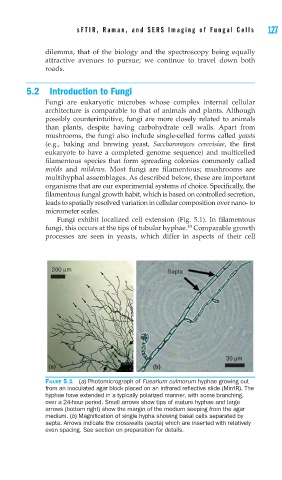
Fungi exhibit localized cell extension (Fig. 5.1). In filamentous
10
fungi, this occurs at the tips of tubular hyphae. Comparable growth
processes are seen in yeasts, which differ in aspects of their cell
FIGURE 5.1 (a) Photomicrograph of Fusarium culmorum hyphae growing out
from an inoculated agar block placed on an infrared refl ective slide (MirrIR). The
hyphae have extended in a typically polarized manner, with some branching,
over a 24-hour period. Small arrows show tips of mature hyphae and large
arrows (bottom right) show the margin of the medium seeping from the agar
medium. (b) Magnifi cation of single hypha showing basal cells separated by
septa. Arrows indicate the crosswalls (septa) which are inserted with relatively
even spacing. See section on preparation for details.