
Page 153 - Vibrational Spectroscopic Imaging for Biomedical Applications
P. 153
sFTIR, Raman, and SERS Imaging of Fungal Cells 129
5.2.1 Specimen Preparation
For informative FTIR, Raman, SERS, and micro-SIMS analysis,
specimens must be chemically pristine, that is, not chemically fixed
or embedded or stained. Our samples are grown in moist cham-
bers across appropriate substrates, nourished from a block of growth
medium. Under these conditions, hyphae will extend 1 to 8 mm
from the block in 24 hours, depending on the species, and their mor-
phology will be indistinguishable from growth on an agar plate.
Extended incubation does not typically lead to ongoing growth, but
can lead to tip morphologies suggestive of stress. Migration of medium
components by capillary action along hyphal walls (Fig. 5.2a, large
arrows) is easily detected. We choose hyphae growing at the colony mar-
gin (Fig. 5.2a, small arrows) since they are more likely to be metaboli-
cally similar. Spores of some plant pathogenic fungi can germinate
on nutrient-free substrates, but this is not generally the case for sap-
rotrophs. Nevertheless, Aspergillus and Neurospora spores have been
shown to germinate at a low frequency without exogenous nutri-
ents, given a humid environment. 8
Sample harvest and preservation must be rapid to prevent cell
degradation or stress-related changes. Our samples are rapidly frozen
by placing them sample side up on a −80ºC metal plate, so that
metabolic processes are arrested within seconds. Frozen samples are
freeze-dried or dried at 37ºC. We find both methods are effective.
Freeze-drying retains cells, three-dimensional structure (good for
scanning electron microscopy) but this can lead to scattering artifacts,
particularly with sFTIR. Air-drying leads to cell collapse, since
16
fungal hyphae are supported by internal hydrostatic pressure acting
against the cell membrane. The membrane is breached by ice crystal
17
damage but the wall restricts the movement of all but small molecules. 18
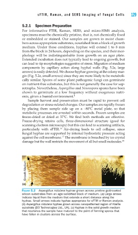
FIGURE 5.2 Aspergillus nidulans hyphae grown across pristine gold-coated
silicon substrates from an agar-solidifi ed block of medium. (a) Large arrows
indicate liquid from the medium that extends a short distance along the
hyphae. Small arrows indicate hyphae appropriate for sFTIR or Raman analysis.
(b) Aspergillus nidulans hyphae grown across nanopatterned region of Klarite
substrate (D3 Technologies Ltd., UK). (c) Hyphae in the solid growth medium
that nourishes the sample have matured to the point of forming spores that
have fallen in clusters across the surface.