
Page 183 - Visions of the Future Chemistry and Life Science
P. 183
164 P. J. KOLSTON
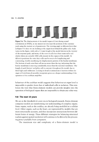
Figure 9.6. The displacement of the model organ of Corti during sound
stimulation at 30kHz, at one instant in time near the position of the response
peak using the normal set of parameters. The viewing angle is different from that
in Figure 9.4. Here we are looking at the organ from behind the pillar cells. Scale
varies in the figure, with only a 1.5-mm length of the model shown in the vicinity
of the response peak, and many of the rows of cells have been removed to aid
clarity (there are actually 1000 cells present in this region of the model). At
different positions the outer hair cells can be seen to be lengthening and
contracting, thereby modifying the displacement pattern of the basilar membrane.
The bottom of each outer hair cell moves more than the top, indicating that the
basilar membrane is moving considerably more than the tectorial membrane. The
length of each Deiters’ and pillar cell is constant throughout the model, due to
their high axial stiffnesses. Looking in detail at animations of motion within the
organ of Corti from all possible viewpoints gives us a deeper understanding of the
operation of the cochlear amplifier.
behaviour of the cochlear model suggests that behaviour at organ level is
impossible to predict from that of individual cells in isolation. This rein-
forces the view that finite-element models can provide insights into the
operation of biological organs that are impossible to obtain any other way.
9.8 The next 10 years
We are at the threshold of a new era in biological research. Finite-element
computer models are transforming our understanding of complete organs.
Some organs, such as the cochlea, are already being modelled at a cellular
level. Other organs, such as the heart, are represented by models that are
more structurally accurate, and they incorporate interactions between dif-
ferent forms of energy. These different strategies for balancing structural
realism against spatial resolution will continue to be driven by the process-
ing power available from computers.
The maximum size and complexity of a finite-element model is