
Page 185 - Visions of the Future Chemistry and Life Science
P. 185
166 P. J. KOLSTON
(a) (b)
(c) (d)
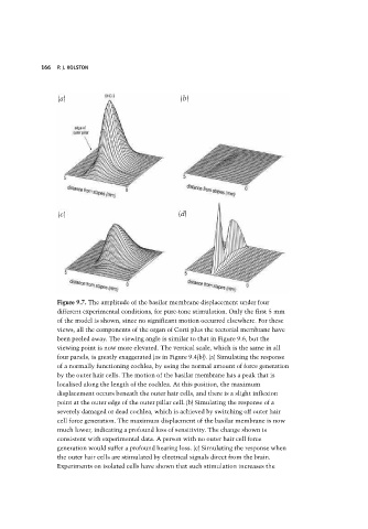
Figure 9.7. The amplitude of the basilar membrane displacement under four
different experimental conditions, for pure-tone stimulation. Only the first 5 mm
of the model is shown, since no significant motion occurred elsewhere. For these
views, all the components of the organ of Corti plus the tectorial membrane have
been peeled away. The viewing angle is similar to that in Figure 9.6, but the
viewing point is now more elevated. The vertical scale, which is the same in all
four panels, is greatly exaggerated (as in Figure 9.4(b)). (a) Simulating the response
of a normally functioning cochlea, by using the normal amount of force generation
by the outer hair cells. The motion of the basilar membrane has a peak that is
localised along the length of the cochlea. At this position, the maximum
displacement occurs beneath the outer hair cells, and there is a slight inflexion
point at the outer edge of the outer pillar cell. (b) Simulating the response of a
severely damaged or dead cochlea, which is achieved by switching off outer hair
cell force generation. The maximum displacment of the basilar membrane is now
much lower, indicating a profound loss of sensitivity. The change shown is
consistent with experimental data. A person with no outer hair cell force
generation would suffer a profound hearing loss. (c) Simulating the response when
the outer hair cells are stimulated by electrical signals direct from the brain.
Experiments on isolated cells have shown that such stimulation increases the