
Page 180 - Visions of the Future Chemistry and Life Science
P. 180
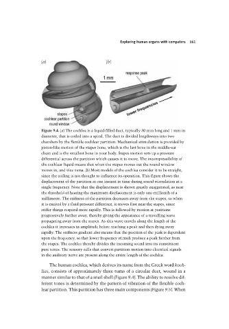
Exploring human organs with computers  161


                                 (a)                        (b)


                                 Figure 9.4. (a) The cochlea is a liquid-filled duct, typically 30 mm long and 1 mm in
                                 diameter, that is coiled into a spiral. The duct is divided lengthways into two
                                 chambers by the flexible cochlear partition. Mechanical stimulation is provided by
                                 piston-like motion of the stapes bone, which is the last bone in the middle-ear
                                 chain and is the smallest bone in your body. Stapes motion sets up a pressure
                                 differential across the partition which causes it to move. The incompressibility of
                                 the cochlear liquid means that when the stapes moves out the round window
                                 moves in, and vice versa. (b) Most models of the cochlea consider it to be straight,
                                 since the coiling is not thought to influence its operation. This figure shows the
                                 displacement of the partition at one instant in time during sound stimulation at a
                                 single frequency. Note that the displacement is shown greatly exaggerated, as near
                                 the threshold of hearing the maximum displacement is only one-millionth of a
                                 millimetre. The stiffness of the partition decreases away from the stapes, so when
                                 it is excited by a fluid pressure difference, it moves first near the stapes, since
                                 stiffer things respond more rapidly. This is followed by motion at positions
                                 progressively further away, thereby giving the appearance of a travelling wave
                                 propagating away from the stapes. As this wave travels along the length of the
                                 cochlea it increases in amplitude before reaching a peak and then dying away
                                 rapidly. The stiffness gradient also means that the position of the peak is dependent
                                 upon the frequency, so that lower frequency stimuli produce a peak further from
                                 the stapes. The cochlea thereby divides the incoming sound into its constituent
                                 pure tones. The sensory cells that convert partition motion into electrical signals
                                 in the auditory nerve are present along the entire length of the cochlea.

                                    The human cochlea, which derives its name from the Greek word koch-
                                 lias, consists of approximately three turns of a circular duct, wound in a
                                 manner similar to that of a snail shell (Figure 9.4). The ability to resolve dif-
                                 ferent tones is determined by the pattern of vibration of the flexible coch-
                                 lear partition. This partition has three main components (Figure 9.5). When