
Page 11 - Advances in Biomechanics and Tissue Regeneration
P. 11
1.3 PATIENT-SPECIFIC GEOMETRY 5
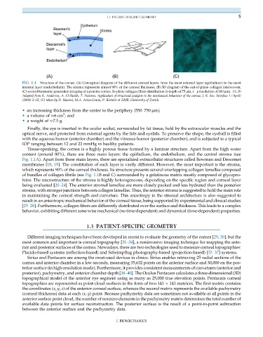
FIG. 1.1 Structure of the cornea. (A) Conceptual diagram of the different corneal layers, from the most external layer (epithelium) to the most
internal layer (endothelium). The stroma represents almost 90% of the corneal thickness; (B) 3D diagram of the out-of-plane collagen interwoven;
(C) second-harmonic generation imaging of a porcine cornea. In-plane collagen fibers distribution (z-depth of 75 μm, x y resolution of 100 μm). (A, B)
Adapted from K. Anderson, A. El-Sheikh, T. Newson, Application of structural analysis to the mechanical behaviour of the cornea, J. R. Soc. Interface 1(April)
(2004) 3–15; (C) taken by D. Haenni, M.A. Ariza-Gracia, P. B€ uchler at ZMB, University of Zurich.
• an increasing thickness from the center to the periphery (550–750 μm);
3
• a volume of 6cm ; and
• a weight of 7.5 g.
Finally, the eye is inserted in the ocular socket, surrounded by fat tissue, held by the extraocular muscles and the
optical nerve, and protected from external agents by the lids and eyelids. To preserve the shape, the eyeball is filled
with the aqueous humor (anterior chamber) and the vitreous humor (posterior chamber), and is subjected to a typical
IOP ranging between 12 and 22 mmHg in healthy patients.
Tissue-speaking, the cornea is a highly porous tissue formed by a laminar structure. Apart from the high water
content (around 80%), there are three main layers: the epithelium, the endothelium, and the central stroma (see
Fig. 1.1A). Apart from these main layers, there are specialized extracellular structures called Bowman and Descemet
membranes [18, 19]. The constitution of each layer is vastly different. However, the most important is the stroma,
which represents 90% of the corneal thickness. Its structure presents several overlapping collagen lamellae composed
of bundles of collagen fibrils (see Fig. 1.1B and C) surrounded by a gelatinous matrix mostly composed of glycopro-
teins. The microstructure of the stroma is highly heterogeneous, depending on the specific region and corneal layer
being evaluated [20–24]. The anterior stromal lamellae are more closely packed and less hydrated than the posterior
stroma, with stronger junctions between collagen lamellas. Thus, the anterior stroma is suggested to hold the main role
in maintaining the corneal strength and curvature. This anisotropy in the stromal architecture is also suggested to
result in an anisotropic mechanical behavior of the corneal tissue, being supported by experimental and clinical studies
[25–28]. Furthermore, collagen fibers are differently distributed over the surface and thickness. This leads to a complex
behavior, exhibiting different zone-wise mechanical (no time-dependent) and dynamical (time-dependent) properties.
1.3 PATIENT-SPECIFIC GEOMETRY
Different imaging techniques have been developed in recent to evaluate the geometry of the cornea [29, 30], but the
most common and important is corneal topography [31–34], a noninvasive imaging technique for mapping the ante-
rior and posterior surfaces of the cornea. Nowadays, there are two technologies used to measure corneal topographies:
Placido-based systems (reflection-based) and Scheimpflug photography-based (projection-based) [35–37] systems.
Sirius and Pentacam are among the most-used devices in clinics. Sirius enables retrieving 25 radial sections of the
cornea and anterior chamber in a few seconds, measuring 35,632 points on the anterior surface and 30,000 on the pos-
terior surface (in high-resolution mode). Furthermore, it provides consistent measurements of curvatures (anterior and
posterior), pachymetry, and anterior chamber depth [38–40]. The Oculus Pentacam calculates a three-dimensional (3D)
topographical model of the anterior eye segment using as many as 25,000 true elevation points. Pentacam corneal
topographies are represented as point cloud surfaces in the form of two 141 141 matrices. The first matrix contains
the coordinates (x, y, z) of the anterior corneal surface, whereas the second matrix represents the available pachymetry
(corneal thickness) data at each (x, y) point. Because pachymetry data are sometimes not available at all points in the
anterior surface point cloud, the number of nonzero elements in the pachymetry matrix determines the total number of
available data points for surface reconstruction. The posterior surface is the result of a point-to-point subtraction
between the anterior surface and the pachymetry data.
I. BIOMECHANICS