
Page 14 - Advances in Biomechanics and Tissue Regeneration
P. 14
8 1. PERSONALIZED CORNEAL BIOMECHANICS
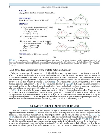
FIG. 1.4 Zero-pressure algorithm. (A) Zero-pressure algorithm accounting for the pull-back algorithm with a consistent mapping of the
fibers onto the current unloaded state; (B) iterative scheme of the algorithm. Adapted from M.Á. Ariza-Gracia, J. Zurita, D.P. Piñero, B. Calvo, J.F. Rodrí-
guez Matas, Automatized patient-specific methodology for numerical determination of biomechanical corneal response, Ann. Biomed. Eng. 44 (5) (2016)
1753–1772.
1.3.3 Stress-Free Configuration of the Eyeball: Reference Geometry
When an eye is measured by a topographer, the identified geometry belongs to a deformed configuration due to the
effect of the IOP (hereafter referred to as the image-based geometry) but the corneal prestress is neglected. Hence, an
accurate stress analysis of the cornea starts by identifying the initial state of stresses due to the physiological IOP pre-
sent on the image-based geometry, or equivalently, the actual geometry associated with the absence of IOP (hereafter
referred to as the zero-pressure geometry), as shown in Fig. 1.4A. Consequently, an iterative algorithm is used to find
the zero-pressure configuration of the eye [43] (see in the algorithm in Fig. 1.4B) that keeps the mesh connectivity
unchanged and iteratively updates the nodal coordinates. Moreover, the local directions of anisotropy (orientation
of collagen fibers) are also consistently pulled back to the current zero-pressure configuration.
In Fig. 1.4,X REF stands for the patient’s geometry reconstructed from the topographer’s data, where X represents an
N n 3 matrix that stores the nodal coordinates of the FE eyeball, with N n the number of nodes in the FE mesh; X k is the
d
zero-pressure configuration identified at iteration k; and X is the deformed configuration obtained when inflating the
k
zero-pressure configuration X k at the IOP pressure. The iterative algorithm updates the zero-pressure geometry, X k ,
d
until the infinite norm of the nodal error between X REF and X is less than a tolerance, TOL.
k
1.4 PATIENT-SPECIFIC MATERIAL BEHAVIOR
A number of material models have been proposed to reproduce the behavior of the cornea, ranging from simple
hyperelastic isotropic materials [44] to more complex models coupling the hyperelastic isotropic response for the
matrix (i.e., neo-Hookean models) with the anisotropic response of the collagen fibers of the eye [7, 25, 41, 42,
45–48]. These material models have been incorporated into computer models of the eye to simulate surgical interven-
tions and tonometry tests in an effort to demonstrate the potential of these in silico models [6, 7, 11, 12, 49–54]. Once a
constitutive model is chosen, it must be particularized for each patient. Unfortunately, many of the methodologies to
retrieve material parameters require a high computational effort and cannot be used in clinics. The proposed predictive
tool relies on a dataset generated by the results of FE simulations of the noncontact tonometry test. The simulations are
I. BIOMECHANICS