
Page 19 - Advances in Biomechanics and Tissue Regeneration
P. 19
1.5 SURGERY SIMULATION 13
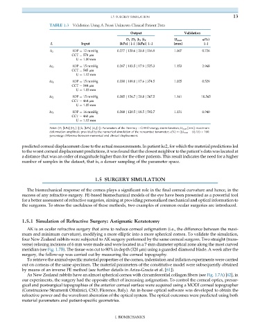
TABLE 1.3 Validation Using A Priori Unknown Clinical Patient Data
Output Validation
D 1 jD 2 jk 1 jk 2 U num e(%)
L Input [kPa] j [–] j [kPa] j [–] [mm] [–]
h 0 IOP ¼ 12 mmHg 0.277 j 120.6 j 20.8 j 516.9 1.007 0.726
CCT ¼ 578 μm
U ¼ 1.00 mm
kc 0 IOP ¼ 15 mmHg 0.267 j 103.5 j 17.9 j 525.3 1.153 2.968
CCT ¼ 545 μm
U ¼ 1.12 mm
kc 1 IOP ¼ 15 mmHg 0.330 j 109.0 j 17.6 j 374.5 1.025 0.529
CCT ¼ 544 μm
U ¼ 1.03 mm
IOP ¼ 15 mmHg 0.385 j 126.7 j 20.8 j 267.5 1.161 10.565
kc 2
CCT ¼ 464 μm
U ¼ 1.05 mm
IOP ¼ 16 mmHg 0.388 j 120.5 j 18.5 j 592.7 1.131 0.940
kc 3
CCT ¼ 460 μm
U ¼ 1.12 mm
Notes:(D 1 [kPa] jD 2 [–] j k 1 [kPa] j k 2 [–]): Parameters of the Demiray + G-H-O energy strain function; U num [mm]: maximum
deformation amplitude provided by the numerical simulation of the noncontact tonometer; E(%) ¼ (jU num Uj/U) 100:
percentage difference between numerical and clinical displacement.
predicted corneal displacement close to the actual measurements. In patient kc2, for which the material predictions led
to the worst corneal displacement predictions, it was found that the closest neighbor to the patient’s data was located at
a distance that was an order of magnitude higher than for the other patients. This result indicates the need for a higher
number of samples in the dataset, that is, a denser sampling of the parameter space.
1.5 SURGERY SIMULATION
The biomechanical response of the cornea plays a significant role in the final corneal curvature and hence, in the
success of any refractive surgery. FE-based biomechanical models of the eye have been presented as a powerful tool
for a better assessment of refractive surgeries, aiming at providing personalized mechanical and optical information to
the surgeons. To stress the usefulness of these methods, two examples of common ocular surgeries are introduced.
1.5.1 Simulation of Refractive Surgery: Astigmatic Keratotomy
AK is an ocular refractive surgery that aims to reduce corneal astigmatism (i.e., the difference between the maxi-
mum and minimum curvature), modifying a more elliptic into a more spherical cornea. To validate the simulation,
four New Zealand rabbits were subjected to AK surgery performed by the same corneal surgeon. Two straight (trans-
verse) relaxing incisions of 6 mm were made and were located in a 7-mm diameter optical zone along the most curved
meridian (see Fig. 1.7B). The tissue was cut to 80% in depth (320 μm) using a guarded diamond blade. A week after the
surgery, the follow-up was carried out by measuring the corneal topography.
To retrieve the animal-specific material properties of the cornea, indentation and inflation experiments were carried
out on corneas of the same specimen. The material parameters of the constitutive model were subsequently obtained
by means of an inverse FE method (see further details in Ariza-Gracia et al. [61]).
As New Zealand rabbits have an almost spherical cornea with circumferential collagen fibers (see Fig. 1.7A)[62], in
our experiments, the surgery had the opposite effect of increasing astigmatism. To control the corneal optics, presur-
gical and postsurgical topographies of the anterior corneal surface were acquired using a MODI corneal topographer
(Construzione Strumenti Oftalmici, CSO, Florence, Italy). An in-house optical software was developed to obtain the
refractive power and the wavefront aberration of the optical system. The optical outcomes were predicted using both
material parameters and patient-specific geometries.
I. BIOMECHANICS