
Page 23 - Advances in Biomechanics and Tissue Regeneration
P. 23
1.6 CONCLUSIONS 17
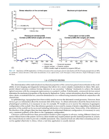
FIG. 1.9 Simulation of ICRS implantation. (A) Stress relaxation at the corneal apex after the introduction of ICRS; (B) main principal stress in MPa;
(C) profile for corneal elevation at the same circumference (5 mm optical zone). (Left) Presurgical corneal elevation. (Right) Postsurgical corneal
elevation.
1.6 CONCLUSIONS
The determination of the individualized mechanical properties of the cornea has gained interest thanks to the avail-
ability of new imaging and diagnostic techniques that allows for a more complex examination in clinics. Still, unex-
pected clinical outcomes continue forcing clinicians to use secondary “refining” treatments to achieve the desired
optical outcomes with the consequent repercussions on a patient’s welfare. Unfortunately, commercial devices cannot
determine the mechanical properties of the stroma by themselves. Today, certain information about the tissue, such as
stress, strain, or material behavior, cannot be determined in vivo without using numerical methods in conjunction with
clinical data.
In ophthalmology, topographers have been widely used to recover the deformed corneal geometry. However, these
devices give no information about the tensional state of the tissue. To obtain information about the tissue behavior in
physiological conditions, it is necessary to use, for example, FE models so as to recover the reference (Lagrangian)
configuration of the eyeball. Once this reference geometry is recovered by means of iterative algorithms, the deformed
geometry of the eyeball can be recovered including information of its tensional state. Noteworthy, if the definition of
the direction of the collagen fibers has been imposed on the deformed (Eulerian) configuration, it is necessary to carry
out a consistent pullback of the fibers to the Lagrangian configuration.
Clinicians could benefit from these personalized models to plan surgeries in advance, testing different clinical sce-
narios without harm to the patient. However, these models may require patient-specific material properties of, at least,
the corneal stroma in order to provide a reliable assessment. Also, numerical tools are often time-consuming and can-
not be applied effectively in clinics, where a real-time application is usually desired.
I. BIOMECHANICS