
Page 28 - Advances in Biomechanics and Tissue Regeneration
P. 28
22 2. BIOMECHANICS OF THE VESTIBULAR SYSTEM: A NUMERICAL SIMULATION
are some authors such as Powers and Howley [7] stating that the receptors inside the vestibular system are sensitive to
any movement change position. The head movements excite the hair cells placed in the sensitive structures, clarified in
the next paragraphs, generating nervous impulses, which are sent to the CNS to recognize the new position.
Patients with vestibular dysfunction exhibit a measurable impairment in motor behavior controlled by the vestib-
ular system (postural control and oculomotor and spatial orientation) and perceptual illusions, such as vertigo [4]. This
and other clinical symptomatology will be detailed in the next section.
The reduced dimensions of such a system are an aspect that has to be reported, since the vestibular system controls
all the balance function comprised in just 8mm.
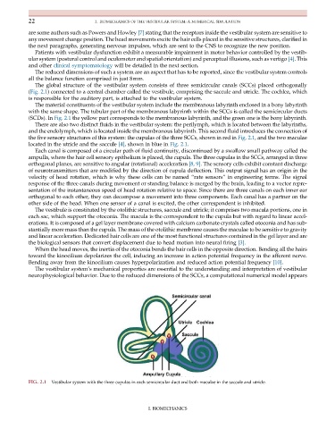
The global structure of the vestibular system consists of three semicircular canals (SCCs) placed orthogonally
(Fig. 2.1) connected to a central chamber called the vestibule, comprising the saccule and utricle. The cochlea, which
is responsible for the auditory part, is attached to the vestibular system.
The material constituents of the vestibular system include the membranous labyrinth enclosed in a bony labyrinth
with the same shape. The tubular part of the membranous labyrinth within the SCCs is called the semicircular ducts
(SCDs). In Fig. 2.1 the yellow part corresponds to the membranous labyrinth, and the green one is the bony labyrinth.
There are also two distinct fluids in the vestibular system: the perilymph, which is located between the labyrinths,
and the endolymph, which is located inside the membranous labyrinth. This second fluid introduces the connection of
the five sensory structures of this system: the cupulas of the three SCCs, shown in red in Fig. 2.1, and the two maculae
located in the utricle and the saccule [4], shown in blue in Fig. 2.1.
Each canal is composed of a circular path of fluid continuity, discontinued by a swallow small pathway called the
ampulla, where the hair cell sensory epithelium is placed, the cupula. The three cupulas in the SCCs, arranged in three
orthogonal planes, are sensitive to angular (rotational) acceleration [8, 9]. The sensory cells exhibit constant discharge
of neurotransmitters that are modified by the direction of cupula deflection. This output signal has an origin in the
velocity of head rotation, which is why these cells can be named “rate sensors” in engineering terms. The signal
response of the three canals during movement or standing balance is merged by the brain, leading to a vector repre-
sentation of the instantaneous speed of head rotation relative to space. Since there are three canals on each inner ear
orthogonal to each other, they can decompose a movement into three components. Each canal has a partner on the
other side of the head. When one sensor of a canal is excited, the other correspondent is inhibited.
The vestibule is constituted by the otolithic structures, saccule and utricle; it comprises two macula portions, one in
each sac, which support the otoconia. The macula is the correspondent to the cupula but with regard to linear accel-
erations. It is composed of a gel layer membrane covered with calcium carbonate crystals called otoconia and has sub-
stantially more mass than the cupula. The mass of the otolithic membrane causes the maculae to be sensitive to gravity
and linear acceleration. Dedicated hair cells are one of the most functional structures contained in the gel layer and are
the biological sensors that convert displacement due to head motion into neural firing [3].
When the head moves, the inertia of the otoconia bends the hair cells in the opposite direction. Bending all the hairs
toward the kinocilium depolarizes the cell, inducing an increase in action potential frequency in the afferent nerve.
Bending away from the kinocilium causes hyperpolarization and reduced action potential frequency [10].
The vestibular system’s mechanical properties are essential to the understanding and interpretation of vestibular
neurophysiological behavior. Due to the reduced dimensions of the SCCs, a computational numerical model appears
FIG. 2.1 Vestibular system with the three cupulas in each semicircular duct and both maculae in the saccule and utricle.
I. BIOMECHANICS