
Page 20 - Advances in Biomechanics and Tissue Regeneration
P. 20
14 1. PERSONALIZED CORNEAL BIOMECHANICS
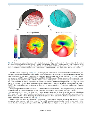
FIG. 1.7 Simulation of astigmatic keratotomy in New Zealand rabbits. (A) Collagen distribution in New Zealand rabbits; (B) FE model of
the surgery (relaxing incisions are outlined in red); (C.1) Sagittal refractive power for specimen 3 measured with MODI; (C.2) Numerical sagittal
refractive power of the FE simulation. Adapted from M.Á. Ariza-Gracia, Á. Ortill es, J.Á. Cristobal, J.F. Rodríguez Matas, B. Calvo, A numerical-experimental
protocol to characterize corneal tissue with an application to predict astigmatic keratotomy surgery, J. Mech. Behav. Biomed. Mater. 74 (2017) 304–314.
From the corneal topography (see Fig. 1.7C), the topographic axis was used to determine the treatment position, and
the topographic cylinder measurement was used to define the length of the incisions. The patient-specific model was
built by transforming a geometrical template into the point cloud of the actual corneal coordinates [12]. The template
was composed of 63,361 nodes (190,083 D.O.F.) and 50,466 C3D8H elements. The chosen mesh is fine enough to prop-
erly capture the contact between the indenter and the cornea and to provide an accurate record of the pressure, area,
and forces measured at the tip. Regarding the boundary conditions, a restrained displacement was imposed at the
scleral rim to mimic the clamping of the sample, and a uniform pressure was imposed on the inner surface to reproduce
the IOP. The contact between the indenter and the cornea was modeled as a frictionless hard contact allowing
separation.
The optical quality of the cornea was used as a criterion to validate the model. Thus, the cylindrical (Cyl) and spher-
ical (Sph) power of the wavefront aberration of the ocular system was used to assess the optical quality.
Optics are purely determined by the geometry of the cornea while geometry depends on IOP and material stiffness
[10]. Hence, only patient-specific models are reliable in terms of refractive outcomes. Although all specimens presented
the same trend, for the sake of simplicity all results correspond to the specimen with the most representative bow-tie
pattern associated with astigmatism (see Fig. 1.7C).
The optical features of the actual geometry are given to a circular area of 3 mm in radius (i.e., the optical zone cor-
responding to the physical pupil of the system). The models are able to reproduce the overall optical quality of the
cornea after the AK surgery. The prediction of the cylindrical power (Cyl) is in good agreement with experiments
I. BIOMECHANICS