
Page 21 - Advances in Biomechanics and Tissue Regeneration
P. 21
1.5 SURGERY SIMULATION 15
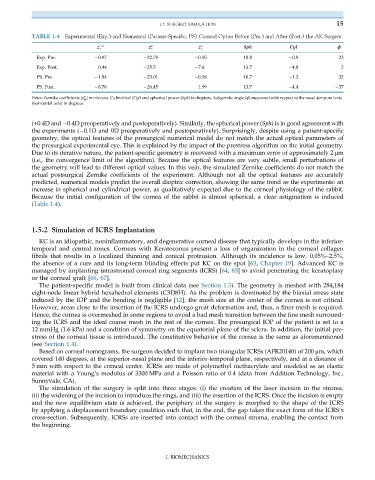
TABLE 1.4 Experimental (Exp.) and Numerical (Patient-Specific, PS) Corneal Optics Before (Pre.) and After (Post.) the AK Surgery
z 2 z 0 2 z 2 2 Sph Cyl ϕ
2
Exp. Pre. 0.97 22.79 0.95 10.8 0.9 23
Exp. Post. 0.44 25.5 7.6 13.7 4.8 2
PS. Pre. 1.94 23.01 0.96 10.7 1.3 32
PS. Post. 6.76 26.45 1.99 13.7 4.4 37
n
Notes: Zernike coefficients (z m ) in microns. Cylindrical (Cyl) and spherical power (Sph) in diopters. Astigmatic angle (ϕ) measured with respect to the nasal-temporal axis
(horizontal axis) in degrees.
(+0.4D and 0.4D preoperatively and postoperatively). Similarly, the spherical power (Sph) is in good agreement with
the experiments ( 0.1D and 0D preoperatively and postoperatively). Surprisingly, despite using a patient-specific
geometry, the optical features of the presurgical numerical model do not match the actual optical parameters of
the presurgical experimental eye. This is explained by the impact of the prestress algorithm on the initial geometry.
Due to its iterative nature, the patient-specific geometry is recovered with a maximum error of approximately 2 μm
(i.e., the convergence limit of the algorithm). Because the optical features are very subtle, small perturbations of
the geometry will lead to different optical values. In this vein, the simulated Zernike coefficients do not match the
actual postsurgical Zernike coefficients of the experiment. Although not all the optical features are accurately
predicted, numerical models predict the overall dioptric correction, showing the same trend as the experiments: an
increase in spherical and cylindrical power, as qualitatively expected due to the corneal physiology of the rabbit.
Because the initial configuration of the cornea of the rabbit is almost spherical, a clear astigmatism is induced
(Table 1.4).
1.5.2 Simulation of ICRS Implantation
KC is an idiopathic, noninflammatory, and degenerative corneal disease that typically develops in the inferior-
temporal and central zones. Corneas with Keratoconus present a loss of organizationinthe cornealcollagen
fibrils that results in a localized thinning and conical protrusion. Although its incidence is low, 0.05%–2.5%,
theabsence ofacureand itslong-term blinding effects put KC on the spot [63, Chapter 19]. Advanced KC is
managed by implanting intrastromal corneal ring segments (ICRS) [64, 65] to avoid penetrating the keratoplasy
or the corneal graft [66, 67].
The patient-specific model is built from clinical data (see Section 1.3). The geometry is meshed with 284,184
eight-node linear hybrid hexahedral elements (C3D8H). As the problem is dominated by the biaxial stress state
induced by the IOP and the bending is negligible [12], the mesh size at the center of the cornea is not critical.
However, areas close to the insertion of the ICRS undergo great deformation and, thus, a finer mesh is required.
Hence, the cornea is overmeshed in some regions to avoid a bad mesh transition between the fine mesh surround-
ing the ICRS and the ideal coarse mesh in the rest of the cornea. The presurgical IOP of the patient is set to a
12 mmHg (1.6 kPa) and a condition of symmetry on the equatorial plane of the sclera. In addition, the initial pre-
stress of the corneal tissue is introduced. The constitutive behavior of the cornea is the same as aforementioned
(see Section 1.4).
Based on corneal nomograms, the surgeon decided to implant two triangular ICRSs (AFR20140) of 200 μm, which
covered 140 degrees, at the superior-nasal plane and the inferior-temporal plane, respectively, and at a distance of
5 mm with respect to the corneal center. ICRSs are made of polymethyl methacrylate and modeled as an elastic
material with a Young’s modulus of 3300 MPa and a Poisson ratio of 0.4 (data from Addition Technology, Inc.,
Sunnyvale, CA).
The simulation of the surgery is split into three stages: (i) the creation of the laser incision in the stroma,
(ii) the widening of the incision to introduce the rings, and (iii) the insertion of the ICRS. Once the incision is empty
and the new equilibrium state is achieved, the periphery of the surgery is morphed to the shape of the ICRS
by applying a displacement boundary condition such that, in the end, the gap takes the exact form of the ICRS’s
cross-section. Subsequently, ICRSs are inserted into contact with the corneal stroma, enabling the contact from
the beginning.
I. BIOMECHANICS