
Page 22 - Advances in Biomechanics and Tissue Regeneration
P. 22
16 1. PERSONALIZED CORNEAL BIOMECHANICS
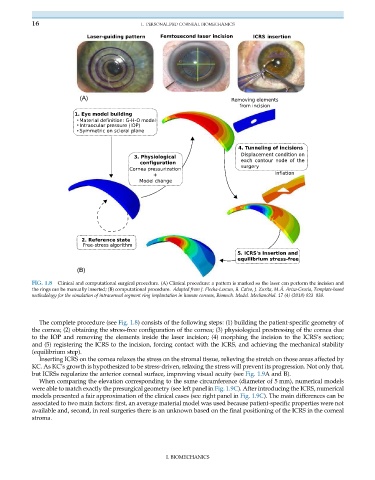
FIG. 1.8 Clinical and computational surgical procedure. (A) Clinical procedure: a pattern is marked so the laser can perform the incision and
the rings can be manually inserted; (B) computational procedure. Adapted from J. Flecha-Lescun, B. Calvo, J. Zurita, M.Á. Ariza-Gracia, Template-based
methodology for the simulation of intracorneal segment ring implantation in human corneas, Biomech. Model. Mechanobiol. 17 (4) (2018) 923–938.
The complete procedure (see Fig. 1.8) consists of the following steps: (1) building the patient-specific geometry of
the cornea; (2) obtaining the stress-free configuration of the cornea; (3) physiological prestressing of the cornea due
to the IOP and removing the elements inside the laser incision; (4) morphing the incision to the ICRS’s section;
and (5) registering the ICRS to the incision, forcing contact with the ICRS, and achieving the mechanical stability
(equilibrium step).
Inserting ICRS on the cornea relaxes the stress on the stromal tissue, relieving the stretch on those areas affected by
KC. As KC’s growth is hypothesized to be stress-driven, relaxing the stress will prevent its progression. Not only that,
but ICRSs regularize the anterior corneal surface, improving visual acuity (see Fig. 1.9A and B).
When comparing the elevation corresponding to the same circumference (diameter of 5 mm), numerical models
were able to match exactly the presurgical geometry (see left panel in Fig. 1.9C). After introducing the ICRS, numerical
models presented a fair approximation of the clinical cases (see right panel in Fig. 1.9C). The main differences can be
associated to two main factors: first, an average material model was used because patient-specific properties were not
available and, second, in real surgeries there is an unknown based on the final positioning of the ICRS in the corneal
stroma.
I. BIOMECHANICS