
Page 12 - Advances in Biomechanics and Tissue Regeneration
P. 12
6 1. PERSONALIZED CORNEAL BIOMECHANICS
The availability of high-resolution topographical data and the patient’s IOP have made it possible to reconstruct a
patient’s specific geometric model of the cornea, which makes it possible to study specific treatments and pathologies,
develop a robust methodology to incorporate a patient’s specific corneal topology into a finite element (FE) model of
the eyeball, and account for the stress-free configuration of the eyeball.
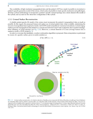
1.3.1 Corneal Surface Reconstruction
A reliable patient-specific FE model of the cornea must incorporate the patient’s topographical data as much as
possible. In this regard, the proposed framework makes use of actual patient data where available, minimizing the
amount of extrapolated data required to build a full 3D FE model amenable for numerical simulations. Current topog-
raphers provide topographical data limited to a corneal area between 8 and 9 mm in diameter due to patient misalign-
ment, blinking, or eyelid aperture (see Fig. 1.2A). However, a corneal diameter of 12 mm (average human size) is
needed to build a 3D FE model [6, 7].
In order to overcome this limitation, a surface continuation algorithm is proposed. Data extrapolation is performed
by means of a quadric surface given in matrix notation as
T
T
x Ax +2B x + c ¼ 0, (1.1)
FIG. 1.2 Corneal surface reconstruction. (A) Anterior elevation of healthy cornea measured with Sirius; (B) surface smoothing at the joint between
the extended surface and the patient’s corneal surface; (C) projection of the 12 mm diameter corneal surface in the optical axis plane. Gray area cor-
responds to the extended surface required in order to achieve a 12-mm diameter (approximating surface). Contour map of the error between the point
cloud data prior and after smoothing (less than 5% at the corneal periphery). Adapted from M.Á. Ariza-Gracia, J. Zurita, D.P. Piñero, B. Calvo, J.F.
Rodríguez Matas, Automatized patient-specific methodology for numerical determination of biomechanical corneal response, Ann. Biomed. Eng. 44 (5) (2016)
1753–1772.
I. BIOMECHANICS