
Page 224 - Advances in Biomechanics and Tissue Regeneration
P. 224
220 11. ANALYSIS OF THE BIOMECHANICAL BEHAVIOR OF INTRAMEDULLARY NAILING
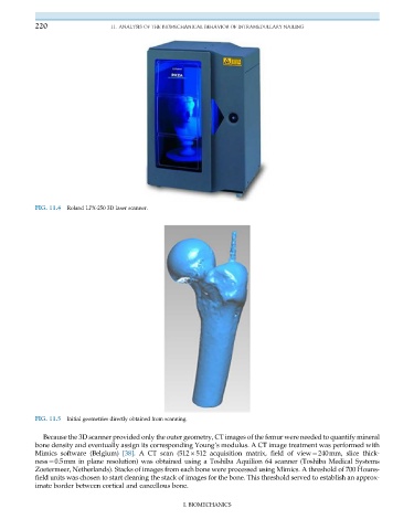
FIG. 11.4 Roland LPX-250 3D laser scanner.
FIG. 11.5 Initial geometries directly obtained from scanning.
Because the 3D scanner provided only the outer geometry, CT images of the femur were needed to quantify mineral
bone density and eventually assign its corresponding Young’s modulus. A CT image treatment was performed with
Mimics software (Belgium) [38]. A CT scan (512 512 acquisition matrix, field of view¼240mm, slice thick-
ness¼0.5mm in plane resolution) was obtained using a Toshiba Aquilion 64 scanner (Toshiba Medical Systems
Zoetermeer, Netherlands). Stacks of images from each bone were processed using Mimics. A threshold of 700 Houns-
field units was chosen to start cleaning the stack of images for the bone. This threshold served to establish an approx-
imate border between cortical and cancellous bone.
I. BIOMECHANICS