
Page 35 - Advances in Biomechanics and Tissue Regeneration
P. 35
2.4 BIOMECHANICAL MODEL OF THE SEMICIRCULAR DUCTS 29
1 2 3 4 5 6 7 8 9 10
FEM
RPIM
NNRPIM
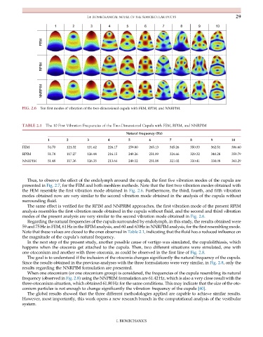
FIG. 2.6 Ten first modes of vibration of the two-dimensional cupula with FEM, RPIM, and NNRPIM.
TABLE 2.1 The 10 First Vibration Frequencies of the Two-Dimensional Cupula with FEM, RPIM, and NNRPIM
Natural frequency (Hz)
1 2 3 4 5 6 7 8 9 10
FEM 54.70 121.32 131.62 226.17 259.80 265.13 345.26 350.33 362.51 386.60
RPIM 51.78 117.27 126.98 216.15 249.26 251.89 324.66 329.32 340.28 359.79
NNRPIM 51.48 117.36 126.35 213.64 249.12 251.08 321.02 330.41 338.08 361.29
Thus, to observe the effect of the endolymph around the cupula, the first five vibration modes of the cupula are
presented in Fig. 2.7, for the FEM and both meshless methods. Note that the first two vibration modes obtained with
the FEM resemble the first vibration mode obtained in Fig. 2.6. Furthermore, the third, fourth, and fifth vibration
modes obtained now are very similar to the second vibration mode obtained in the analysis of the cupula without
surrounding fluid.
The same effect is verified for the RPIM and NNPRIM approaches, the first vibration mode of the present RPIM
analysis resembles the first vibration mode obtained in the cupula without fluid, and the second and third vibration
modes of the present analysis are very similar to the second vibration mode obtained in Fig. 2.6.
Regarding the natural frequencies of the cupula surrounded by endolymph, in this study, the results obtained were
59 and 75Hz in FEM, 61Hz in the RPIM analysis, and 60 and 63Hz in NNRPIM analysis, for the first resembling mode.
Note that these values are closed to the ones observed in Table 2.1, indicating that the fluid has a reduced influence on
the magnitude of the cupula’s natural frequency.
In the next step of the present study, another possible cause of vertigo was simulated, the cupulolithiasis, which
happens when the otoconia get attached to the cupula. Then, two different situations were simulated, one with
one otoconium and another with three otoconia, as could be observed in the first line of Fig. 2.8.
The goal is to understand if the inclusion of the otoconia changes significantly the natural frequency of the cupula.
Since the results obtained in the previous analyses with the three formulations were very similar, in Fig. 2.8, only the
results regarding the NNRPIM formulation are presented.
When one otoconium (or one otoconium group) is considered, the frequencies of the cupula resembling its natural
frequency (observed in Fig. 2.8) using the NNPRIM formulation are 61.42Hz, which is also a very close result with the
three-otoconium situation, which obtained 61.80Hz for the same conditions. This may indicate that the size of the oto-
conium particles is not enough to change significantly the vibration frequency of the cupula [40].
The global results showed that the three different methodologies applied are capable to achieve similar results.
However, most importantly, this work opens a new research branch in the computational analysis of the vestibular
system.
I. BIOMECHANICS