
Page 142 - Bio Engineering Approaches to Cancer Diagnosis and Treatment
P. 142
140 CHAPTER 6 Laser-assisted cancer treatment
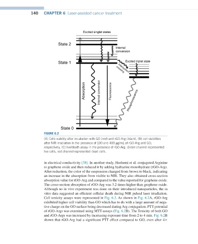
FIGURE 6.2
(A) Cells viability after incubation with GO (red) and rGO-Arg (black), (B) cell viabilities
after NIR irradiation in the presence of 100 and 400 µg/mL of rGO-Arg and GO,
respectively, (C) live/death assay in the presence of rGO-Arg. Green channel represented
live cells, red channel represented dead cells.
in electrical conductivity [38]. In another study, Hashemi et al. conjugated Arginine
to graphene oxide and then reduced it by adding hydrazine monohydrate (rGO-Arg).
After reduction, the color of the suspension changed from brown to black, indicating
an increase in the absorption from visible to NIR. They also obtained cross-section
absorption value for rGO-Arg and compared to the value reported for graphene oxide.
The cross-section absorption of rGO-Arg was 3.2 times higher than graphene oxide.
Although no in vivo experiment was done on their introduced nanoparticles, the in
vitro data suggested an efficient cellular death during NIR pulsed laser irradiation.
Cell toxicity assays were represented in Fig. 6.2. As shown in Fig. 6.2A, rGO-Arg
exhibited higher cell viability than GO which has to do with a large amount of nega-
tive charge on the GO surface being decreased during Arg conjugation. PTT potential
of rGO-Args was examined using MTT-assays (Fig. 6.2B). The Toxicity of both GO
and rGO-Args was increased by increasing exposure time from 2 to 4 min. Fig. 6.2B
shown that rGO-Arg had a significant PTT effect compared to GO, even after 4×